 Movie
Movie Controller

[English] 日本語
 Yorodumi
- PDB-8d79: Crystal structure of a four-tetrad, parallel, and Na+ stabilized ... -
+ Open data

- Basic information
| Entry | Database: PDB / ID: 8d79 | ||||||||||||||||||||||||||||
|---|---|---|---|---|---|---|---|---|---|---|---|---|---|---|---|---|---|---|---|---|---|---|---|---|---|---|---|---|---|
| Title | Crystal structure of a four-tetrad, parallel, and Na+ stabilized Tetrahymena thermophila telomeric G-quadruplex in complex with N-methyl mesoporphyrin IX | ||||||||||||||||||||||||||||
 Components Components | DNA (5'-D(* Keywords KeywordsDNA / G-quadruplex / Parallel / four-tetrad | Function / homology | N-METHYLMESOPORPHYRIN / DNA / DNA (> 10) |  Function and homology information Function and homology informationBiological species |   Tetrahymena thermophila (eukaryote) Tetrahymena thermophila (eukaryote)Method |  X-RAY DIFFRACTION / SYNCHROTRON / MOLECULAR REPLACEMENT / Resolution: 1.99 Å X-RAY DIFFRACTION / SYNCHROTRON / MOLECULAR REPLACEMENT / Resolution: 1.99 Å  Authors AuthorsChen, E.V. / Beseiso, D. / Yatsunyk, L.A. | Funding support | |  United States, 1items United States, 1items
 CitationJournal: To Be Published CitationJournal: To Be PublishedTitle: Biophysical and structural characterization of telomeric G-quadruplexes from Tetrahymena thermophila in complex with N-Methyl Mesoporphyrin IX Authors: Beseiso, D. / Chen, E.V. / Yatsunyk, L.A. History |
|
- Structure visualization
| Structure viewer | Molecule: MolmilJmol/JSmol |
|---|
- Downloads & links
-Download
| PDBx/mmCIF format | 8d79.cif.gz | 48.1 KB | Display | PDBx/mmCIF format |
|---|---|---|---|---|
| PDB format | pdb8d79.ent.gz | 28.3 KB | Display | PDB format |
| PDBx/mmJSON format | 8d79.json.gz | Tree view | PDBx/mmJSON format | |
| Others |  Other downloads Other downloads |
-Validation report
| Arichive directory | https://data.pdbj.org/pub/pdb/validation_reports/d7/8d79ftp://data.pdbj.org/pub/pdb/validation_reports/d7/8d79 | HTTPS FTP |
|---|
-Related structure data
| Related structure data |  8d78C  6w9pS S: Starting model for refinement C: citing same article ( |
|---|---|
| Similar structure data |
-Links
 PDBj
PDBj


- Assembly
| Deposited unit | 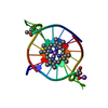
| ||||||||||||
|---|---|---|---|---|---|---|---|---|---|---|---|---|---|
| 1 |
| ||||||||||||
| Unit cell |
| ||||||||||||
| Components on special symmetry positions |
|
-Components
| #1: DNA chain | Mass: 7655.879 Da / Num. of mol.: 1 / Source method: obtained synthetically / Source: (synth.) Tetrahymena thermophila (eukaryote) | ||||||||||
|---|---|---|---|---|---|---|---|---|---|---|---|
| #2: Chemical | ChemComp-NA /   Mass: 22.990 Da / Num. of mol.: 5 / Source method: obtained synthetically / Formula: Na Mass: 22.990 Da / Num. of mol.: 5 / Source method: obtained synthetically / Formula: Na#3: Chemical | ChemComp-MMP / |   Mass: 580.716 Da / Num. of mol.: 1 / Source method: obtained synthetically / Formula: C35H40N4O4 Mass: 580.716 Da / Num. of mol.: 1 / Source method: obtained synthetically / Formula: C35H40N4O4#4: Chemical |   Mass: 118.174 Da / Num. of mol.: 2 / Source method: obtained synthetically / Formula: C6H14O2 / Comment: precipitant*YM Mass: 118.174 Da / Num. of mol.: 2 / Source method: obtained synthetically / Formula: C6H14O2 / Comment: precipitant*YM#5: Water | ChemComp-HOH / |  Mass: 18.015 Da / Num. of mol.: 12 / Source method: isolated from a natural source / Formula: H2O Mass: 18.015 Da / Num. of mol.: 12 / Source method: isolated from a natural source / Formula: H2OHas ligand of interest | N | Nonpolymer details | The authors state that the MPD ligands modelled in the structure are shown to have LINKS to the ...The authors state that the MPD ligands modelled in the structure are shown to have LINKS to the main DNA chain. Rather than removing the MPD to eliminate the LINKS, they have preserved the MPDs in the structure as a part of the crystal packing interaction. It should be noted that the MPD molecules are at symmetry-positions and have a high amount of disorder. | |
-Experimental details
-Experiment
| Experiment | Method: X-RAY DIFFRACTION / Number of used crystals: 1 |
|---|
- Sample preparation
| Crystal | Density Matthews: 1.93 Å3/Da / Density % sol: 34.56 % |
|---|---|
| Crystal grow | Temperature: 298 K / Method: vapor diffusion, hanging drop Details: Well condition contained 100 mM NaCl, 100 mM NaI, 100 mM NaF, 30% MPD, and 50 mM Na cacodylate pH 6.5 |
-Data collection
| Diffraction | Mean temperature: 196 K / Serial crystal experiment: N |
|---|---|
| Diffraction source | Source: SYNCHROTRON / Site: APS / Beamline: 24-ID-C / Wavelength: 0.97918 Å |
| Detector | Type: DECTRIS EIGER X 16M / Detector: PIXEL / Date: Feb 12, 2021 |
| Radiation | Protocol: SINGLE WAVELENGTH / Monochromatic (M) / Laue (L): M / Scattering type: x-ray |
| Radiation wavelength | Wavelength: 0.97918 Å / Relative weight: 1 |
| Reflection | Resolution: 1.98→32.97 Å / Num. obs: 3905 / % possible obs: 96.2 % / Redundancy: 3.3 % / Biso Wilson estimate: 52.83 Å2 / CC1/2: 0.987 / Rmerge(I) obs: 0.118 / Rpim(I) all: 0.075 / Rrim(I) all: 0.14 / Net I/σ(I): 9.5 |
| Reflection shell | Resolution: 1.98→2.03 Å / Redundancy: 2.8 % / Rmerge(I) obs: 0.846 / Mean I/σ(I) obs: 0.8 / Num. unique obs: 689 / CC1/2: 0.836 / Rpim(I) all: 0.599 / % possible all: 73.1 |
- Processing
| Software |
| ||||||||||||||||||||||||||||||||||||||||
|---|---|---|---|---|---|---|---|---|---|---|---|---|---|---|---|---|---|---|---|---|---|---|---|---|---|---|---|---|---|---|---|---|---|---|---|---|---|---|---|---|---|
| Refinement | Method to determine structure: MOLECULAR REPLACEMENT Starting model: 6W9P Resolution: 1.99→32.97 Å / SU ML: 0 / Cross valid method: FREE R-VALUE / σ(F): 1.37 / Phase error: 31.4912 Stereochemistry target values: GeoStd + Monomer Library + CDL v1.2
| ||||||||||||||||||||||||||||||||||||||||
| Solvent computation | Shrinkage radii: 0.9 Å / VDW probe radii: 1.11 Å / Solvent model: FLAT BULK SOLVENT MODEL | ||||||||||||||||||||||||||||||||||||||||
| Displacement parameters | Biso mean: 77.61 Å2 | ||||||||||||||||||||||||||||||||||||||||
| Refinement step | Cycle: LAST / Resolution: 1.99→32.97 Å
| ||||||||||||||||||||||||||||||||||||||||
| Refine LS restraints |
| ||||||||||||||||||||||||||||||||||||||||
| LS refinement shell | Resolution: 1.99→32.97 Å
| ||||||||||||||||||||||||||||||||||||||||
| Refinement TLS params. | Method: refined / Origin x: -4.19725759583 Å / Origin y: 7.41567938218 Å / Origin z: 9.3528971543 Å
| ||||||||||||||||||||||||||||||||||||||||
| Refinement TLS group | Selection details: all |
