 Movie
Movie Controller

[English] 日本語
 Yorodumi
- PDB-8d6n: Q108K:K40L:T53A:R58L:Q38F:Q4F mutant of hCRBPII bound to syntheti... -
+ Open data

- Basic information
| Entry | Database: PDB / ID: 8d6n | ||||||
|---|---|---|---|---|---|---|---|
| Title | Q108K:K40L:T53A:R58L:Q38F:Q4F mutant of hCRBPII bound to synthetic fluorophore CM1V | ||||||
 Components Components | Retinol-binding protein 2 | ||||||
 Keywords Keywords | TRANSPORT PROTEIN / hCRBPII | ||||||
| Function / homology |  Function and homology information Function and homology informationvitamin A metabolic process / molecular carrier activity / retinoid binding / retinal binding / retinol binding / epidermis development / fatty acid transport / Retinoid metabolism and transport / fatty acid binding / nucleus / cytosol Similarity search - Function | ||||||
| Biological species |  Homo sapiens (human) Homo sapiens (human) | ||||||
| Method |  X-RAY DIFFRACTION / SYNCHROTRON / MOLECULAR REPLACEMENT / Resolution: 1.42 Å X-RAY DIFFRACTION / SYNCHROTRON / MOLECULAR REPLACEMENT / Resolution: 1.42 Å | ||||||
 Authors Authors | Bingham, C.R. / Geiger, J.H. / Borhan, B. | ||||||
| Funding support |  United States, 1items United States, 1items
| ||||||
 Citation Citation | Journal: Analyst / Year: 2023 Title: Light controlled reversible Michael addition of cysteine: a new tool for dynamic site-specific labeling of proteins. Authors: Maity, S. / Bingham, C. / Sheng, W. / Ehyaei, N. / Chakraborty, D. / Tahmasebi-Nick, S. / Kimmel, T.E. / Vasileiou, C. / Geiger, J.H. / Borhan, B. | ||||||
| History |
|
- Structure visualization
| Structure viewer | Molecule: MolmilJmol/JSmol |
|---|
- Downloads & links
-Download
| PDBx/mmCIF format | 8d6n.cif.gz | 47.4 KB | Display | PDBx/mmCIF format |
|---|---|---|---|---|
| PDB format | pdb8d6n.ent.gz | 30.9 KB | Display | PDB format |
| PDBx/mmJSON format | 8d6n.json.gz | Tree view | PDBx/mmJSON format | |
| Others |  Other downloads Other downloads |
-Validation report
| Summary document | 8d6n_validation.pdf.gz | 678.3 KB | Display | wwPDB validaton report |
|---|---|---|---|---|
| Full document | 8d6n_full_validation.pdf.gz | 681 KB | Display | |
| Data in XML | 8d6n_validation.xml.gz | 9.3 KB | Display | |
| Data in CIF | 8d6n_validation.cif.gz | 11.5 KB | Display | |
| Arichive directory | https://data.pdbj.org/pub/pdb/validation_reports/d6/8d6nftp://data.pdbj.org/pub/pdb/validation_reports/d6/8d6n | HTTPS FTP |
-Related structure data
| Related structure data | 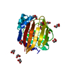 8d6hC  8d6lC  8db2C 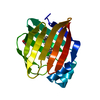 8dn1C  4qypS S: Starting model for refinement C: citing same article ( |
|---|---|
| Similar structure data |
-Links
 PDBj
PDBj


- Assembly
| Deposited unit | 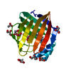
| ||||||||||||
|---|---|---|---|---|---|---|---|---|---|---|---|---|---|
| 1 |
| ||||||||||||
| Unit cell |
|
-Components
-Protein , 1 types, 1 molecules A
| #1: Protein | Mass: 15546.509 Da / Num. of mol.: 1 / Mutation: Q108K, K40L, T53A, R58L, Q38F, Q4F Source method: isolated from a genetically manipulated source Source: (gene. exp.) Homo sapiens (human) / Gene: RBP2, CRBP2 / Production host:  |
|---|
-Non-polymers , 5 types, 56 molecules 
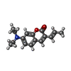


| #2: Chemical |  Mass: 59.044 Da / Num. of mol.: 3 / Source method: obtained synthetically / Formula: C2H3O2 Mass: 59.044 Da / Num. of mol.: 3 / Source method: obtained synthetically / Formula: C2H3O2#3: Chemical | ChemComp-RH6 / ( |  Mass: 257.328 Da / Num. of mol.: 1 / Source method: obtained synthetically / Formula: C16H19NO2 Mass: 257.328 Da / Num. of mol.: 1 / Source method: obtained synthetically / Formula: C16H19NO2#4: Chemical | ChemComp-GOL /  Mass: 92.094 Da / Num. of mol.: 7 / Source method: obtained synthetically / Formula: C3H8O3 Mass: 92.094 Da / Num. of mol.: 7 / Source method: obtained synthetically / Formula: C3H8O3#5: Chemical |  Mass: 104.061 Da / Num. of mol.: 2 / Source method: obtained synthetically / Formula: C3H4O4 Mass: 104.061 Da / Num. of mol.: 2 / Source method: obtained synthetically / Formula: C3H4O4#6: Water | ChemComp-HOH / | Mass: 18.015 Da / Num. of mol.: 43 / Source method: isolated from a natural source / Formula: H2O |
|---|
-Details
| Has ligand of interest | Y |
|---|---|
| Has protein modification | Y |
-Experimental details
-Experiment
| Experiment | Method: X-RAY DIFFRACTION / Number of used crystals: 1 |
|---|
- Sample preparation
| Crystal | Density Matthews: 2.04 Å3/Da / Density % sol: 39.77 % |
|---|---|
| Crystal grow | Temperature: 297 K / Method: vapor diffusion, hanging drop / pH: 4 Details: PEG 4000, Ammonium Acetate, Sodium Acetate, pH 4.0 - 4.8 PH range: pH 4.0 -4.8 |
-Data collection
| Diffraction | Mean temperature: 100 K / Serial crystal experiment: N |
|---|---|
| Diffraction source | Source: SYNCHROTRON / Site: APS / Beamline: 21-ID-D / Wavelength: 1.1272 Å |
| Detector | Type: DECTRIS EIGER X 9M / Detector: PIXEL / Date: Jul 23, 2021 |
| Radiation | Protocol: SINGLE WAVELENGTH / Monochromatic (M) / Laue (L): M / Scattering type: x-ray |
| Radiation wavelength | Wavelength: 1.1272 Å / Relative weight: 1 |
| Reflection | Resolution: 1.42→33.55 Å / Num. obs: 22948 / % possible obs: 97.28 % / Redundancy: 6.4 % / Biso Wilson estimate: 16.18 Å2 / Rpim(I) all: 0.014 / Rrim(I) all: 0.036 / Net I/σ(I): 22949 |
| Reflection shell | Resolution: 1.42→1.47 Å / Num. unique obs: 2284 / CC1/2: 0.987 / Rpim(I) all: 0.079 / Rrim(I) all: 0.198 / % possible all: 96.21 |
- Processing
| Software |
| |||||||||||||||||||||||||||||||||||||||||||||||||||||||||||||||||||||||||||||||||||||||||||||||||||||||||
|---|---|---|---|---|---|---|---|---|---|---|---|---|---|---|---|---|---|---|---|---|---|---|---|---|---|---|---|---|---|---|---|---|---|---|---|---|---|---|---|---|---|---|---|---|---|---|---|---|---|---|---|---|---|---|---|---|---|---|---|---|---|---|---|---|---|---|---|---|---|---|---|---|---|---|---|---|---|---|---|---|---|---|---|---|---|---|---|---|---|---|---|---|---|---|---|---|---|---|---|---|---|---|---|---|---|---|
| Refinement | Method to determine structure: MOLECULAR REPLACEMENT Starting model: 4QYP Resolution: 1.42→33.55 Å / SU ML: 0.166 / Cross valid method: FREE R-VALUE / σ(F): 1.43 / Phase error: 25.0478 Stereochemistry target values: GeoStd + Monomer Library + CDL v1.2
| |||||||||||||||||||||||||||||||||||||||||||||||||||||||||||||||||||||||||||||||||||||||||||||||||||||||||
| Solvent computation | Shrinkage radii: 0.9 Å / VDW probe radii: 1.11 Å / Solvent model: FLAT BULK SOLVENT MODEL | |||||||||||||||||||||||||||||||||||||||||||||||||||||||||||||||||||||||||||||||||||||||||||||||||||||||||
| Displacement parameters | Biso mean: 21.5 Å2 | |||||||||||||||||||||||||||||||||||||||||||||||||||||||||||||||||||||||||||||||||||||||||||||||||||||||||
| Refinement step | Cycle: LAST / Resolution: 1.42→33.55 Å
| |||||||||||||||||||||||||||||||||||||||||||||||||||||||||||||||||||||||||||||||||||||||||||||||||||||||||
| Refine LS restraints |
| |||||||||||||||||||||||||||||||||||||||||||||||||||||||||||||||||||||||||||||||||||||||||||||||||||||||||
| LS refinement shell |
|
