neuronal stem cell division / regulation of leukocyte cell-cell adhesion / regulation of leukocyte tethering or rolling / alpha-(1->3)-fucosyltransferase activity / 4-galactosyl-N-acetylglucosaminide 3-alpha-L-fucosyltransferase / 4-galactosyl-N-acetylglucosaminide 3-alpha-L-fucosyltransferase activity / Lewis x epitope biosynthetic process / Lewis blood group biosynthesis / glycosphingolipid biosynthetic process / fucosyltransferase activity ...neuronal stem cell division / regulation of leukocyte cell-cell adhesion / regulation of leukocyte tethering or rolling / alpha-(1->3)-fucosyltransferase activity / 4-galactosyl-N-acetylglucosaminide 3-alpha-L-fucosyltransferase / 4-galactosyl-N-acetylglucosaminide 3-alpha-L-fucosyltransferase activity / Lewis x epitope biosynthetic process / Lewis blood group biosynthesis / glycosphingolipid biosynthetic process / fucosyltransferase activity / oligosaccharide biosynthetic process / L-fucose catabolic process / N-glycan processing / protein O-linked glycosylation / glycoprotein biosynthetic process / protein N-linked glycosylation / polysaccharide biosynthetic process / trans-Golgi network membrane / trans-Golgi network / positive regulation of neuron projection development / neuron differentiation / carbohydrate metabolic process / Golgi membrane / Golgi apparatus / protein homodimerization activity Similarity search - Function
In the structure databanks used in Yorodumi, some data are registered as the other names, "COVID-19 virus" and "2019-nCoV". Here are the details of the virus and the list of structure data.
Jan 31, 2019. EMDB accession codes are about to change! (news from PDBe EMDB page)
EMDB accession codes are about to change! (news from PDBe EMDB page)
The allocation of 4 digits for EMDB accession codes will soon come to an end. Whilst these codes will remain in use, new EMDB accession codes will include an additional digit and will expand incrementally as the available range of codes is exhausted. The current 4-digit format prefixed with “EMD-” (i.e. EMD-XXXX) will advance to a 5-digit format (i.e. EMD-XXXXX), and so on. It is currently estimated that the 4-digit codes will be depleted around Spring 2019, at which point the 5-digit format will come into force.
The EM Navigator/Yorodumi systems omit the EMD- prefix.
Related info.:Q: What is EMD? / ID/Accession-code notation in Yorodumi/EM Navigator
Yorodumi is a browser for structure data from EMDB, PDB, SASBDB, etc.
This page is also the successor to EM Navigator detail page, and also detail information page/front-end page for Omokage search.
The word "yorodu" (or yorozu) is an old Japanese word meaning "ten thousand". "mi" (miru) is to see.
Related info.:EMDB / PDB / SASBDB / Comparison of 3 databanks / Yorodumi Search / Aug 31, 2016. New EM Navigator & Yorodumi / Yorodumi Papers / Jmol/JSmol / Function and homology information / Changes in new EM Navigator and Yorodumi
 Movie
Movie Controller


 Open data
 Basic information
 Components
 Keywords
 Function and homology information
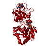
 Homo sapiens (human)
 X-RAY DIFFRACTION /
  Authors
 United States, 1items
  Citation
 Structure visualization
 Downloads & links
 Other downloads


 PDBj
PDBj Assembly
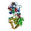
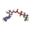


 Mass: 643.313 Da / Num. of mol.: 1 / Source method: obtained synthetically / Formula: C16H22F3N5O15P2 / Feature type: SUBJECT OF INVESTIGATION
 Type: RNA linking / Mass: 443.201 Da / Num. of mol.: 1 / Source method: obtained synthetically / Formula: C10H15N5O11P2 / Feature type: SUBJECT OF INVESTIGATION / Comment: GDP, energy-carrying molecule*YM
 Mass: 96.063 Da / Num. of mol.: 2 / Source method: obtained synthetically / Formula: SO4
 Sample preparation
 Processing