 Movie
Movie Controller


+ Open data

- Basic information
| Entry | Database: PDB / ID: 8d09 | ||||||
|---|---|---|---|---|---|---|---|
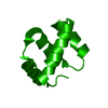
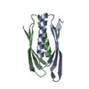
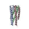
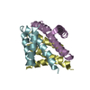
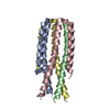
| Title | Hallucinated C4 protein assembly HALC4_136 | ||||||
 Components Components | HALC4_136 | ||||||
 Keywords Keywords | DE NOVO PROTEIN / De novo design Hallucination Cyclic Oligomer ProteinMPNN | ||||||
| Biological species | synthetic construct (others) | ||||||
| Method |  X-RAY DIFFRACTION / SYNCHROTRON / MOLECULAR REPLACEMENT / Resolution: 1.9 Å X-RAY DIFFRACTION / SYNCHROTRON / MOLECULAR REPLACEMENT / Resolution: 1.9 Å | ||||||
 Authors Authors | Ragotte, R.J. / Bera, A.K. / Wicky, B.I.M. / Milles, L.F. / Baker, D. | ||||||
| Funding support |  United States, 1items United States, 1items
| ||||||
 Citation Citation | Journal: Science / Year: 2022 Title: Hallucinating symmetric protein assemblies. Authors: B I M Wicky / L F Milles / A Courbet / R J Ragotte / J Dauparas / E Kinfu / S Tipps / R D Kibler / M Baek / F DiMaio / X Li / L Carter / A Kang / H Nguyen / A K Bera / D Baker / Abstract: Deep learning generative approaches provide an opportunity to broadly explore protein structure space beyond the sequences and structures of natural proteins. Here, we use deep network hallucination ...Deep learning generative approaches provide an opportunity to broadly explore protein structure space beyond the sequences and structures of natural proteins. Here, we use deep network hallucination to generate a wide range of symmetric protein homo-oligomers given only a specification of the number of protomers and the protomer length. Crystal structures of seven designs are very similar to the computational models (median root mean square deviation: 0.6 angstroms), as are three cryo-electron microscopy structures of giant 10-nanometer rings with up to 1550 residues and symmetry; all differ considerably from previously solved structures. Our results highlight the rich diversity of new protein structures that can be generated using deep learning and pave the way for the design of increasingly complex components for nanomachines and biomaterials. | ||||||
| History |
|
- Structure visualization
| Structure viewer | Molecule:  MolmilJmol/JSmol MolmilJmol/JSmol |
|---|
- Downloads & links
-Download
| PDBx/mmCIF format | 8d09.cif.gz | 41.8 KB | Display | PDBx/mmCIF format |
|---|---|---|---|---|
| PDB format | pdb8d09.ent.gz | 25.4 KB | Display | PDB format |
| PDBx/mmJSON format | 8d09.json.gz | Tree view | PDBx/mmJSON format | |
| Others |  Other downloads Other downloads |
-Validation report
| Arichive directory | https://data.pdbj.org/pub/pdb/validation_reports/d0/8d09ftp://data.pdbj.org/pub/pdb/validation_reports/d0/8d09 | HTTPS FTP |
|---|
-Related structure data


-Links
 PDBj
PDBj
- Assembly
| Deposited unit | 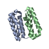
| ||||||||||||
|---|---|---|---|---|---|---|---|---|---|---|---|---|---|
| 1 | 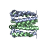
| ||||||||||||
| Unit cell |
|
-Components
| #1: Protein | Mass: 7608.969 Da / Num. of mol.: 2 Source method: isolated from a genetically manipulated source Source: (gene. exp.) synthetic construct (others) / Production host:  #2: Water | ChemComp-HOH / |  Mass: 18.015 Da / Num. of mol.: 36 / Source method: isolated from a natural source / Formula: H2O Mass: 18.015 Da / Num. of mol.: 36 / Source method: isolated from a natural source / Formula: H2O |
|---|
-Experimental details
-Experiment
| Experiment | Method: X-RAY DIFFRACTION / Number of used crystals: 1 |
|---|
- Sample preparation
| Crystal | Density Matthews: 1.78 Å3/Da / Density % sol: 31.08 % |
|---|---|
| Crystal grow | Temperature: 293 K / Method: vapor diffusion, sitting drop / pH: 5 / Details: 0.1M SPG buffer pH 5 25% w/v PEG 1500 |
-Data collection
| Diffraction | Mean temperature: 100 K / Serial crystal experiment: N |
|---|---|
| Diffraction source | Source: SYNCHROTRON / Site: APS / Beamline: 24-ID-C / Wavelength: 0.97918 Å |
| Detector | Type: DECTRIS EIGER2 X 16M / Detector: PIXEL / Date: Mar 13, 2022 |
| Radiation | Protocol: SINGLE WAVELENGTH / Monochromatic (M) / Laue (L): M / Scattering type: x-ray |
| Radiation wavelength | Wavelength: 0.97918 Å / Relative weight: 1 |
| Reflection | Resolution: 1.9→31.34 Å / Num. obs: 8869 / % possible obs: 99.52 % / Redundancy: 13.1 % / Biso Wilson estimate: 37.36 Å2 / CC1/2: 0.981 / Net I/σ(I): 9.45 |
| Reflection shell | Resolution: 1.9→1.968 Å / Mean I/σ(I) obs: 3.95 / Num. unique obs: 875 / CC1/2: 0.832 / % possible all: 100 |
- Processing
| Software |
| ||||||||||||||||||||||||||||
|---|---|---|---|---|---|---|---|---|---|---|---|---|---|---|---|---|---|---|---|---|---|---|---|---|---|---|---|---|---|
| Refinement | Method to determine structure: MOLECULAR REPLACEMENT Starting model: Design model Resolution: 1.9→31.34 Å / SU ML: 0.1581 / Cross valid method: FREE R-VALUE / σ(F): 1.4 / Phase error: 31.1739 Stereochemistry target values: GeoStd + Monomer Library + CDL v1.2
| ||||||||||||||||||||||||||||
| Solvent computation | Shrinkage radii: 0.9 Å / VDW probe radii: 1.1 Å / Solvent model: FLAT BULK SOLVENT MODEL | ||||||||||||||||||||||||||||
| Displacement parameters | Biso mean: 37.36 Å2 | ||||||||||||||||||||||||||||
| Refinement step | Cycle: LAST / Resolution: 1.9→31.34 Å
| ||||||||||||||||||||||||||||
| Refine LS restraints |
| ||||||||||||||||||||||||||||
| LS refinement shell |
|
