 Movie
Movie Controller


+ Open data

- Basic information
| Entry | Database: PDB / ID: 8d00 | ||||||
|---|---|---|---|---|---|---|---|
| Title | Structure of the Arabidopsis thaliana SPIRAL2 TOG domain | ||||||
 Components Components | Microtubule-associated protein TORTIFOLIA1 | ||||||
 Keywords Keywords | STRUCTURAL PROTEIN / TOG / HEAT / Alpha-solenoid / Microtubule-binding | ||||||
| Function / homology |  Function and homology information Function and homology informationcortical microtubule, transverse to long axis / circumnutation / unidimensional cell growth / microtubule binding Similarity search - Function | ||||||
| Biological species |  | ||||||
| Method |  X-RAY DIFFRACTION / SYNCHROTRON / MOLECULAR REPLACEMENT / Resolution: 2.8 Å X-RAY DIFFRACTION / SYNCHROTRON / MOLECULAR REPLACEMENT / Resolution: 2.8 Å | ||||||
 Authors Authors | Slep, K.C. / Bolhuis, D.L. | ||||||
| Funding support |  United States, 1items United States, 1items
| ||||||
 Citation Citation | Journal: Plant Cell / Year: 2023 Title: A structurally divergent TOG domain stabilizes microtubule minus ends in Arabidopsis Authors: Fan, Y. / Bilkey, N. / Bolhuis, D.L. / Slep, K.C. / Dixit, R. | ||||||
| History |
|
- Structure visualization
| Structure viewer | Molecule: MolmilJmol/JSmol |
|---|
- Downloads & links
-Download
| PDBx/mmCIF format | 8d00.cif.gz | 140.4 KB | Display | PDBx/mmCIF format |
|---|---|---|---|---|
| PDB format | pdb8d00.ent.gz | 96.7 KB | Display | PDB format |
| PDBx/mmJSON format | 8d00.json.gz | Tree view | PDBx/mmJSON format | |
| Others |  Other downloads Other downloads |
-Validation report
| Summary document | 8d00_validation.pdf.gz | 423 KB | Display | wwPDB validaton report |
|---|---|---|---|---|
| Full document | 8d00_full_validation.pdf.gz | 425.2 KB | Display | |
| Data in XML | 8d00_validation.xml.gz | 11.9 KB | Display | |
| Data in CIF | 8d00_validation.cif.gz | 15 KB | Display | |
| Arichive directory | https://data.pdbj.org/pub/pdb/validation_reports/d0/8d00ftp://data.pdbj.org/pub/pdb/validation_reports/d0/8d00 | HTTPS FTP |
-Related structure data
| Similar structure data |
|---|
-Links
 PDBj
PDBj- Assembly
| Deposited unit | 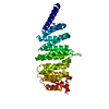
| ||||||||||||
|---|---|---|---|---|---|---|---|---|---|---|---|---|---|
| 1 |
| ||||||||||||
| Unit cell |
|
-Components
| #1: Protein | Mass: 32920.035 Da / Num. of mol.: 1 Source method: isolated from a genetically manipulated source Source: (gene. exp.)  |
|---|---|
| #2: Chemical | ChemComp-IOD /   Mass: 126.904 Da / Num. of mol.: 1 / Source method: obtained synthetically / Formula: I Mass: 126.904 Da / Num. of mol.: 1 / Source method: obtained synthetically / Formula: I |
| #3: Water | ChemComp-HOH /  Mass: 18.015 Da / Num. of mol.: 7 / Source method: isolated from a natural source / Formula: H2O Mass: 18.015 Da / Num. of mol.: 7 / Source method: isolated from a natural source / Formula: H2O |
| Has ligand of interest | N |
-Experimental details
-Experiment
| Experiment | Method: X-RAY DIFFRACTION / Number of used crystals: 1 |
|---|
- Sample preparation
| Crystal | Density Matthews: 2.88 Å3/Da / Density % sol: 57.25 % / Description: rods 50 x 50 x 400 microns |
|---|---|
| Crystal grow | Temperature: 293 K / Method: vapor diffusion, hanging drop Details: 2 ul 15.7 mg/mL SPIRAL2 TOG domain (residues 33-333) protein + 2 ul of a 1 ml well solution (27.5% w/v PEG3350, 150 mM ammonium phosphate, 280 mM sodium iodide) |
-Data collection
| Diffraction | Mean temperature: 100 K / Serial crystal experiment: N |
|---|---|
| Diffraction source | Source: SYNCHROTRON / Site: APS / Beamline: 22-ID / Wavelength: 1 Å |
| Detector | Type: DECTRIS EIGER X 16M / Detector: PIXEL / Date: Feb 9, 2021 |
| Radiation | Monochromator: Si 111. Rosenbaum-Rock double-crystal monochromator: liquid nitrogen cooled; sagitally focusing 2nd crystal, Rosenbaum-Rock vertical focusing mirror Protocol: SINGLE WAVELENGTH / Monochromatic (M) / Laue (L): M / Scattering type: x-ray |
| Radiation wavelength | Wavelength: 1 Å / Relative weight: 1 |
| Reflection | Resolution: 2.8→50 Å / Num. obs: 9490 / % possible obs: 98.7 % / Redundancy: 11.5 % / Biso Wilson estimate: 58.9 Å2 / CC1/2: 0.993 / CC star: 0.998 / Rsym value: 0.164 / Net I/σ(I): 19.7 |
| Reflection shell | Resolution: 2.8→2.9 Å / Mean I/σ(I) obs: 2.6 / Num. unique obs: 944 / CC1/2: 0.872 / CC star: 0.965 / Rsym value: 0.842 |
- Processing
| Software |
| ||||||||||||||||||||||||||||||||||||||||||||||||||||||||
|---|---|---|---|---|---|---|---|---|---|---|---|---|---|---|---|---|---|---|---|---|---|---|---|---|---|---|---|---|---|---|---|---|---|---|---|---|---|---|---|---|---|---|---|---|---|---|---|---|---|---|---|---|---|---|---|---|---|
| Refinement | Method to determine structure: MOLECULAR REPLACEMENT Starting model: AlphaFold prediction Resolution: 2.8→38.36 Å / SU ML: 0.3475 / Cross valid method: FREE R-VALUE / σ(F): 0.17 / Phase error: 26.7654 Stereochemistry target values: GeoStd + Monomer Library + CDL v1.2
| ||||||||||||||||||||||||||||||||||||||||||||||||||||||||
| Solvent computation | Shrinkage radii: 0.9 Å / VDW probe radii: 1.1 Å / Solvent model: FLAT BULK SOLVENT MODEL | ||||||||||||||||||||||||||||||||||||||||||||||||||||||||
| Displacement parameters | Biso mean: 66.99 Å2 | ||||||||||||||||||||||||||||||||||||||||||||||||||||||||
| Refinement step | Cycle: LAST / Resolution: 2.8→38.36 Å
| ||||||||||||||||||||||||||||||||||||||||||||||||||||||||
| Refine LS restraints |
| ||||||||||||||||||||||||||||||||||||||||||||||||||||||||
| LS refinement shell |
| ||||||||||||||||||||||||||||||||||||||||||||||||||||||||
| Refinement TLS params. | Method: refined / Origin x: -9.79538249122 Å / Origin y: -5.82707630509 Å / Origin z: -41.1953209771 Å
| ||||||||||||||||||||||||||||||||||||||||||||||||||||||||
| Refinement TLS group | Selection details: all |
