 Movie
Movie Controller


+ Open data

- Basic information
| Entry | Database: PDB / ID: 8cxk | ||||||
|---|---|---|---|---|---|---|---|
| Title | Structure of the C. elegans HIM-3 R93Y mutant | ||||||
 Components Components | HORMA domain-containing protein | ||||||
 Keywords Keywords | PEPTIDE BINDING PROTEIN / meiotic HORMAD / PCH-2 / HORMA domain / Hop1 | ||||||
| Function / homology |  Function and homology information Function and homology informationregulation of centriole-centriole cohesion / meiotic chromosome segregation / synaptonemal complex assembly / homologous chromosome segregation / homologous chromosome pairing at meiosis / synaptonemal complex / lateral element / reciprocal meiotic recombination / sister chromatid cohesion / condensed nuclear chromosome ...regulation of centriole-centriole cohesion / meiotic chromosome segregation / synaptonemal complex assembly / homologous chromosome segregation / homologous chromosome pairing at meiosis / synaptonemal complex / lateral element / reciprocal meiotic recombination / sister chromatid cohesion / condensed nuclear chromosome / chromosome / DNA binding Similarity search - Function | ||||||
| Biological species |  | ||||||
| Method |  X-RAY DIFFRACTION / SYNCHROTRON / MOLECULAR REPLACEMENT / Resolution: 1.3 Å X-RAY DIFFRACTION / SYNCHROTRON / MOLECULAR REPLACEMENT / Resolution: 1.3 Å | ||||||
 Authors Authors | Ego, K.M. / Russo, A. / Giacopazzi, S. / Deshong, A. / Menon, M. / Ortiz, V. / Bhalla, N. / Corbett, K.D. | ||||||
| Funding support |  United States, 1items United States, 1items
| ||||||
 Citation Citation | Journal: Plos Genet. / Year: 2023 Title: The conserved AAA ATPase PCH-2 distributes its regulation of meiotic prophase events through multiple meiotic HORMADs in C. elegans. Authors: Russo, A.E. / Giacopazzi, S. / Deshong, A. / Menon, M. / Ortiz, V. / Ego, K.M. / Corbett, K.D. / Bhalla, N. | ||||||
| History |
|
- Structure visualization
| Structure viewer | Molecule: MolmilJmol/JSmol |
|---|
- Downloads & links
-Download
| PDBx/mmCIF format | 8cxk.cif.gz | 189.1 KB | Display | PDBx/mmCIF format |
|---|---|---|---|---|
| PDB format | pdb8cxk.ent.gz | 125.2 KB | Display | PDB format |
| PDBx/mmJSON format | 8cxk.json.gz | Tree view | PDBx/mmJSON format | |
| Others |  Other downloads Other downloads |
-Validation report
| Arichive directory | https://data.pdbj.org/pub/pdb/validation_reports/cx/8cxkftp://data.pdbj.org/pub/pdb/validation_reports/cx/8cxk | HTTPS FTP |
|---|
-Related structure data
| Related structure data |  4trkS S: Starting model for refinement |
|---|---|
| Similar structure data | |
| Experimental dataset #1 | Data set type: diffraction image data / Metadata reference: 10.15785/SBGRID/910 |
-Links
 PDBj
PDBj
- Assembly
| Deposited unit | 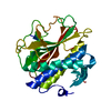
| ||||||||||||
|---|---|---|---|---|---|---|---|---|---|---|---|---|---|
| 1 |
| ||||||||||||
| Unit cell |
|
-Components
| #1: Protein | Mass: 33172.809 Da / Num. of mol.: 1 / Mutation: R93Y Source method: isolated from a genetically manipulated source Source: (gene. exp.)  |
|---|---|
| #2: Water | ChemComp-HOH /  Mass: 18.015 Da / Num. of mol.: 252 / Source method: isolated from a natural source / Formula: H2O Mass: 18.015 Da / Num. of mol.: 252 / Source method: isolated from a natural source / Formula: H2O |
-Experimental details
-Experiment
| Experiment | Method: X-RAY DIFFRACTION / Number of used crystals: 1 |
|---|
- Sample preparation
| Crystal | Density Matthews: 2.59 Å3/Da / Density % sol: 52.44 % |
|---|---|
| Crystal grow | Temperature: 293 K / Method: vapor diffusion, hanging drop / Details: 1.4-1.6 M sodium malonate pH 6.5-7.0 |
-Data collection
| Diffraction | Mean temperature: 100 K / Serial crystal experiment: N |
|---|---|
| Diffraction source | Source: SYNCHROTRON / Site: ALS / Beamline: 8.3.1 / Wavelength: 1.1158 Å |
| Detector | Type: DECTRIS PILATUS 6M / Detector: PIXEL / Date: Sep 14, 2018 |
| Radiation | Protocol: SINGLE WAVELENGTH / Monochromatic (M) / Laue (L): M / Scattering type: x-ray |
| Radiation wavelength | Wavelength: 1.1158 Å / Relative weight: 1 |
| Reflection | Resolution: 1.3→70 Å / Num. obs: 82280 / % possible obs: 100 % / Redundancy: 19.8 % / Biso Wilson estimate: 19.36 Å2 / CC1/2: 1 / Rmerge(I) obs: 0.054 / Rrim(I) all: 0.056 / Net I/σ(I): 27.38 |
| Reflection shell | Resolution: 1.3→1.38 Å / Redundancy: 19.7 % / Rmerge(I) obs: 2.027 / Num. unique obs: 13303 / CC1/2: 0.677 / Rrim(I) all: 2.081 / % possible all: 100 |
- Processing
| Software |
| |||||||||||||||||||||||||||||||||||||||||||||||||||||||||||||||||||||||||||||||||||||||||||||||||||||||||||||||||||||||||||||||||||||||||||||||||||||||||||||||||||||||||||||||||||||||||||||||||||||||||||||||||||||||||
|---|---|---|---|---|---|---|---|---|---|---|---|---|---|---|---|---|---|---|---|---|---|---|---|---|---|---|---|---|---|---|---|---|---|---|---|---|---|---|---|---|---|---|---|---|---|---|---|---|---|---|---|---|---|---|---|---|---|---|---|---|---|---|---|---|---|---|---|---|---|---|---|---|---|---|---|---|---|---|---|---|---|---|---|---|---|---|---|---|---|---|---|---|---|---|---|---|---|---|---|---|---|---|---|---|---|---|---|---|---|---|---|---|---|---|---|---|---|---|---|---|---|---|---|---|---|---|---|---|---|---|---|---|---|---|---|---|---|---|---|---|---|---|---|---|---|---|---|---|---|---|---|---|---|---|---|---|---|---|---|---|---|---|---|---|---|---|---|---|---|---|---|---|---|---|---|---|---|---|---|---|---|---|---|---|---|---|---|---|---|---|---|---|---|---|---|---|---|---|---|---|---|---|---|---|---|---|---|---|---|---|---|---|---|---|---|---|---|---|
| Refinement | Method to determine structure: MOLECULAR REPLACEMENT Starting model: 4TRK Resolution: 1.3→63.42 Å / SU ML: 0.1672 / Cross valid method: FREE R-VALUE / σ(F): 1.36 / Phase error: 15.794 Stereochemistry target values: GeoStd + Monomer Library + CDL v1.2
| |||||||||||||||||||||||||||||||||||||||||||||||||||||||||||||||||||||||||||||||||||||||||||||||||||||||||||||||||||||||||||||||||||||||||||||||||||||||||||||||||||||||||||||||||||||||||||||||||||||||||||||||||||||||||
| Solvent computation | Shrinkage radii: 0.9 Å / VDW probe radii: 1.11 Å / Solvent model: FLAT BULK SOLVENT MODEL | |||||||||||||||||||||||||||||||||||||||||||||||||||||||||||||||||||||||||||||||||||||||||||||||||||||||||||||||||||||||||||||||||||||||||||||||||||||||||||||||||||||||||||||||||||||||||||||||||||||||||||||||||||||||||
| Displacement parameters | Biso mean: 29.1 Å2 | |||||||||||||||||||||||||||||||||||||||||||||||||||||||||||||||||||||||||||||||||||||||||||||||||||||||||||||||||||||||||||||||||||||||||||||||||||||||||||||||||||||||||||||||||||||||||||||||||||||||||||||||||||||||||
| Refinement step | Cycle: LAST / Resolution: 1.3→63.42 Å
| |||||||||||||||||||||||||||||||||||||||||||||||||||||||||||||||||||||||||||||||||||||||||||||||||||||||||||||||||||||||||||||||||||||||||||||||||||||||||||||||||||||||||||||||||||||||||||||||||||||||||||||||||||||||||
| Refine LS restraints |
| |||||||||||||||||||||||||||||||||||||||||||||||||||||||||||||||||||||||||||||||||||||||||||||||||||||||||||||||||||||||||||||||||||||||||||||||||||||||||||||||||||||||||||||||||||||||||||||||||||||||||||||||||||||||||
| LS refinement shell |
|
