 Movie
Movie Controller

[English] 日本語
 Yorodumi
- PDB-8cwp: X-ray crystal structure of NTHi Protein D bound to a putative gly... -
+ Open data

- Basic information
| Entry | Database: PDB / ID: 8cwp | ||||||
|---|---|---|---|---|---|---|---|
| Title | X-ray crystal structure of NTHi Protein D bound to a putative glycerol moiety | ||||||
 Components Components | Glycerophosphoryl diester phosphodiesterase | ||||||
 Keywords Keywords | HYDROLASE / NTHi / otitis media / outer membrane protein / phosphodiesterase / lipo-glycerophosphodiesterase / sn-glycero-3-phosphocholine / choline / sn-glycerol 3-phosphate | ||||||
| Function / homology | Glycerophosphodiester phosphodiesterase domain / Glycerophosphoryl diester phosphodiesterase family / GP-PDE domain profile. / PLC-like phosphodiesterase, TIM beta/alpha-barrel domain superfamily / phosphoric diester hydrolase activity / lipid metabolic process / Glycerophosphoryl diester phosphodiesterase Function and homology information Function and homology information | ||||||
| Biological species |  Haemophilus influenzae (bacteria) Haemophilus influenzae (bacteria) | ||||||
| Method |  X-RAY DIFFRACTION / SYNCHROTRON / MOLECULAR REPLACEMENT / Resolution: 1.8 Å X-RAY DIFFRACTION / SYNCHROTRON / MOLECULAR REPLACEMENT / Resolution: 1.8 Å | ||||||
 Authors Authors | Jones, S.P. / Cook, K.H. / Holmquist, M.L. / Almekinder, L. / DeLaney, A. / Labbe, N. / Perdue, J. / Jackson, N. / Charles, R. / Pichichero, M. ...Jones, S.P. / Cook, K.H. / Holmquist, M.L. / Almekinder, L. / DeLaney, A. / Labbe, N. / Perdue, J. / Jackson, N. / Charles, R. / Pichichero, M. / Kaur, R. / Michel, L. / Gleghorn, M.L. | ||||||
| Funding support |  United States, 1items United States, 1items
| ||||||
 Citation Citation | Journal: Proteins / Year: 2023 Title: Vaccine target and carrier molecule nontypeable Haemophilus influenzae protein D dimerizes like the close Escherichia coli GlpQ homolog but unlike other known homolog dimers. Authors: Jones, S.P. / Cook, K.H. / Holmquist, M.L. / Almekinder, L.J. / Delaney, A.M. / Charles, R. / Labbe, N. / Perdue, J. / Jackson, N. / Pichichero, M.E. / Kaur, R. / Michel, L.V. / Gleghorn, M.L. | ||||||
| History |
|
- Structure visualization
| Structure viewer | Molecule: MolmilJmol/JSmol |
|---|
- Downloads & links
-Download
| PDBx/mmCIF format | 8cwp.cif.gz | 276 KB | Display | PDBx/mmCIF format |
|---|---|---|---|---|
| PDB format | pdb8cwp.ent.gz | 185.5 KB | Display | PDB format |
| PDBx/mmJSON format | 8cwp.json.gz | Tree view | PDBx/mmJSON format | |
| Others |  Other downloads Other downloads |
-Validation report
| Summary document | 8cwp_validation.pdf.gz | 1.1 MB | Display | wwPDB validaton report |
|---|---|---|---|---|
| Full document | 8cwp_full_validation.pdf.gz | 1.1 MB | Display | |
| Data in XML | 8cwp_validation.xml.gz | 18.7 KB | Display | |
| Data in CIF | 8cwp_validation.cif.gz | 29.2 KB | Display | |
| Arichive directory | https://data.pdbj.org/pub/pdb/validation_reports/cw/8cwpftp://data.pdbj.org/pub/pdb/validation_reports/cw/8cwp | HTTPS FTP |
-Related structure data
| Related structure data | 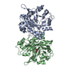 1ydyS S: Starting model for refinement |
|---|---|
| Similar structure data |
-Links
 PDBj
PDBj- Assembly
| Deposited unit | 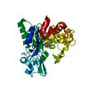
| ||||||||||||
|---|---|---|---|---|---|---|---|---|---|---|---|---|---|
| 1 | 
| ||||||||||||
| Unit cell |
| ||||||||||||
| Components on special symmetry positions |
|
-Components
| #1: Protein | Mass: 41278.863 Da / Num. of mol.: 1 Source method: isolated from a genetically manipulated source Source: (gene. exp.) Haemophilus influenzae (bacteria) / Gene: glpQ, CHBNIII1_03270 / Production host: |
|---|---|
| #2: Chemical | ChemComp-NA /   Mass: 22.990 Da / Num. of mol.: 1 / Source method: obtained synthetically / Formula: Na Mass: 22.990 Da / Num. of mol.: 1 / Source method: obtained synthetically / Formula: Na |
| #3: Chemical | ChemComp-GOL /   Mass: 92.094 Da / Num. of mol.: 1 / Source method: obtained synthetically / Formula: C3H8O3 / Feature type: SUBJECT OF INVESTIGATION Mass: 92.094 Da / Num. of mol.: 1 / Source method: obtained synthetically / Formula: C3H8O3 / Feature type: SUBJECT OF INVESTIGATION |
| #4: Water | ChemComp-HOH /  Mass: 18.015 Da / Num. of mol.: 457 / Source method: isolated from a natural source / Formula: H2O Mass: 18.015 Da / Num. of mol.: 457 / Source method: isolated from a natural source / Formula: H2O |
| Has ligand of interest | Y |
-Experimental details
-Experiment
| Experiment | Method: X-RAY DIFFRACTION / Number of used crystals: 1 |
|---|
- Sample preparation
| Crystal | Density Matthews: 2.73 Å3/Da / Density % sol: 55 % Description: The crystals were approximately 3 millimeters x 120 micrometers (Visually appeared thinner in the third dimension). |
|---|---|
| Crystal grow | Temperature: 293.15 K / Method: vapor diffusion, hanging drop / pH: 5.5 / Details: 20% w/v PEG3000, 0.1 M Sodium Citrate pH 5.5 |
-Data collection
| Diffraction | Mean temperature: 100 K Ambient temp details: Cryostream (Exact temperature unknown) Serial crystal experiment: N |
|---|---|
| Diffraction source | Source: SYNCHROTRON / Site: NSLS-II / Beamline: 17-ID-2 / Wavelength: 0.97927 Å |
| Detector | Type: DECTRIS EIGER X 16M / Detector: PIXEL / Date: Jul 12, 2021 |
| Radiation | Protocol: SINGLE WAVELENGTH / Monochromatic (M) / Laue (L): M / Scattering type: x-ray |
| Radiation wavelength | Wavelength: 0.97927 Å / Relative weight: 1 |
| Reflection | Resolution: 1.8→28 Å / Num. obs: 41845 / % possible obs: 99.7 % / Redundancy: 14.5 % / Biso Wilson estimate: 37.1 Å2 / CC1/2: 0.999 / Rmerge(I) obs: 0.08 / Net I/σ(I): 17.5 |
| Reflection shell | Resolution: 1.8→1.85 Å / Redundancy: 13.6 % / Rmerge(I) obs: 2.6 / Mean I/σ(I) obs: 0.98 / Num. unique obs: 2934 / CC1/2: 0.394 / % possible all: 96 |
- Processing
| Software |
| |||||||||||||||||||||||||||||||||||||||||||||||||||||||||||||||||||||||||||||||||||||||||||||||||||||||||
|---|---|---|---|---|---|---|---|---|---|---|---|---|---|---|---|---|---|---|---|---|---|---|---|---|---|---|---|---|---|---|---|---|---|---|---|---|---|---|---|---|---|---|---|---|---|---|---|---|---|---|---|---|---|---|---|---|---|---|---|---|---|---|---|---|---|---|---|---|---|---|---|---|---|---|---|---|---|---|---|---|---|---|---|---|---|---|---|---|---|---|---|---|---|---|---|---|---|---|---|---|---|---|---|---|---|---|
| Refinement | Method to determine structure: MOLECULAR REPLACEMENT Starting model: 1YDY Resolution: 1.8→28 Å / SU ML: 0.2342 / Cross valid method: FREE R-VALUE / σ(F): 1.33 / Phase error: 19.878 Stereochemistry target values: GeoStd + Monomer Library + CDL v1.2
| |||||||||||||||||||||||||||||||||||||||||||||||||||||||||||||||||||||||||||||||||||||||||||||||||||||||||
| Solvent computation | Shrinkage radii: 0.9 Å / VDW probe radii: 1.11 Å / Solvent model: FLAT BULK SOLVENT MODEL | |||||||||||||||||||||||||||||||||||||||||||||||||||||||||||||||||||||||||||||||||||||||||||||||||||||||||
| Displacement parameters | Biso mean: 44.24 Å2 | |||||||||||||||||||||||||||||||||||||||||||||||||||||||||||||||||||||||||||||||||||||||||||||||||||||||||
| Refinement step | Cycle: LAST / Resolution: 1.8→28 Å
| |||||||||||||||||||||||||||||||||||||||||||||||||||||||||||||||||||||||||||||||||||||||||||||||||||||||||
| Refine LS restraints |
| |||||||||||||||||||||||||||||||||||||||||||||||||||||||||||||||||||||||||||||||||||||||||||||||||||||||||
| LS refinement shell |
| |||||||||||||||||||||||||||||||||||||||||||||||||||||||||||||||||||||||||||||||||||||||||||||||||||||||||
| Refinement TLS params. | Method: refined / Origin x: -0.855320487543 Å / Origin y: -10.7265812324 Å / Origin z: -22.8939257229 Å
| |||||||||||||||||||||||||||||||||||||||||||||||||||||||||||||||||||||||||||||||||||||||||||||||||||||||||
| Refinement TLS group | Selection details: all |
