[English] 日本語
Yorodumi
- PDB-8cwi: Fab arm of antibody 10G4 bound to CoV-2 receptor binding domain (RBD) -

+
Open data


ID or keywords:

Loading...

-
Basic information

Entry
Database: PDB / ID: 8cwi
TitleFab arm of antibody 10G4 bound to CoV-2 receptor binding domain (RBD)
Components
  • Heavy chain of Fab arm of antibody 10G4
  • Light chain of Fab arm of antibody 10G4
  • Spike protein S1
KeywordsIMMUNE SYSTEM / antibody / CoV-2 / receptor binding domain / class 5 epitope
Function / homology
Function and homology information


Maturation of spike protein / viral translation / Translation of Structural Proteins / Virion Assembly and Release / host cell surface / host extracellular space / suppression by virus of host tetherin activity / Induction of Cell-Cell Fusion / structural constituent of virion / entry receptor-mediated virion attachment to host cell ...Maturation of spike protein / viral translation / Translation of Structural Proteins / Virion Assembly and Release / host cell surface / host extracellular space / suppression by virus of host tetherin activity / Induction of Cell-Cell Fusion / structural constituent of virion / entry receptor-mediated virion attachment to host cell / host cell endoplasmic reticulum-Golgi intermediate compartment membrane / receptor-mediated endocytosis of virus by host cell / membrane fusion / Attachment and Entry / positive regulation of viral entry into host cell / receptor-mediated virion attachment to host cell / receptor ligand activity / host cell surface receptor binding / fusion of virus membrane with host plasma membrane / fusion of virus membrane with host endosome membrane / viral envelope / virion attachment to host cell / SARS-CoV-2 activates/modulates innate and adaptive immune responses / host cell plasma membrane / virion membrane / identical protein binding / membrane / plasma membrane
Similarity search - Function
Spike (S) protein S1 subunit, receptor-binding domain, SARS-CoV-2 / Spike (S) protein S1 subunit, N-terminal domain, SARS-CoV-like / Betacoronavirus spike (S) glycoprotein S1 subunit N-terminal (NTD) domain profile. / Spike glycoprotein, N-terminal domain superfamily / Betacoronavirus spike (S) glycoprotein S1 subunit C-terminal (CTD) domain profile. / Spike glycoprotein, betacoronavirus / Spike (S) protein S1 subunit, receptor-binding domain, betacoronavirus / Spike S1 subunit, receptor binding domain superfamily, betacoronavirus / Betacoronavirus spike glycoprotein S1, receptor binding / Spike glycoprotein S1, N-terminal domain, betacoronavirus-like ...Spike (S) protein S1 subunit, receptor-binding domain, SARS-CoV-2 / Spike (S) protein S1 subunit, N-terminal domain, SARS-CoV-like / Betacoronavirus spike (S) glycoprotein S1 subunit N-terminal (NTD) domain profile. / Spike glycoprotein, N-terminal domain superfamily / Betacoronavirus spike (S) glycoprotein S1 subunit C-terminal (CTD) domain profile. / Spike glycoprotein, betacoronavirus / Spike (S) protein S1 subunit, receptor-binding domain, betacoronavirus / Spike S1 subunit, receptor binding domain superfamily, betacoronavirus / Betacoronavirus spike glycoprotein S1, receptor binding / Spike glycoprotein S1, N-terminal domain, betacoronavirus-like / Betacoronavirus-like spike glycoprotein S1, N-terminal / Spike glycoprotein S2, coronavirus, heptad repeat 1 / Spike glycoprotein S2, coronavirus, heptad repeat 2 / Coronavirus spike (S) glycoprotein S2 subunit heptad repeat 2 (HR2) region profile. / Coronavirus spike (S) glycoprotein S2 subunit heptad repeat 1 (HR1) region profile. / Spike glycoprotein S2 superfamily, coronavirus / Spike glycoprotein S2, coronavirus / Coronavirus spike glycoprotein S2 / Coronavirus spike glycoprotein S1, C-terminal / Coronavirus spike glycoprotein S1, C-terminal
Similarity search - Domain/homology
Biological speciesSevere acute respiratory syndrome coronavirus 2
Homo sapiens (human)
MethodX-RAY DIFFRACTION / SYNCHROTRON / MOLECULAR REPLACEMENT / molecular replacement / Resolution: 1.873 Å
AuthorsLangley, D.B. / Christ, D.
Funding support Australia, 1items
OrganizationGrant numberCountry
National Health and Medical Research Council (NHMRC, Australia) Australia
CitationJournal: To Be Published
Title: Neutralization of CoV-2 omicron lineages by affinity-matured class 5 antibodies
Authors: Langley, D.B. / Christ, D.
History
DepositionMay 19, 2022Deposition site: RCSB / Processing site: RCSB
Revision 1.0May 24, 2023Provider: repository / Type: Initial release
Revision 1.1Apr 3, 2024Group: Data collection / Refinement description
Category: chem_comp_atom / chem_comp_bond / pdbx_initial_refinement_model

-
Structure visualization

Structure viewerMolecule:
MolmilJmol/JSmol

Downloads & links

-
Assembly

Deposited unit
A: Spike protein S1
H: Heavy chain of Fab arm of antibody 10G4
L: Light chain of Fab arm of antibody 10G4
hetero molecules


Theoretical massNumber of molelcules
Total (without water)72,82510
Polymers71,0423
Non-polymers1,7827
Water12,016667
1


  • Idetical with deposited unit
  • defined by author&software
TypeNameSymmetry operationNumber
identity operation1_555x,y,z1
Buried area7240 Å2
ΔGint-44 kcal/mol
Surface area29460 Å2
MethodPISA
Unit cell
Length a, b, c (Å)53.691, 88.639, 81.398
Angle α, β, γ (deg.)90.000, 95.030, 90.000
Int Tables number4
Space group name H-MP1211

-
Components

-
Protein , 1 types, 1 molecules A

#1: Protein Spike protein S1


Mass: 22975.688 Da / Num. of mol.: 1 / Fragment: receptor binding domain (RBD)
Source method: isolated from a genetically manipulated source
Source: (gene. exp.) Severe acute respiratory syndrome coronavirus 2
Gene: S, 2 / Cell line (production host): ExpiCHO / Production host: 
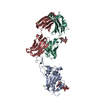
Cricetulus griseus (Chinese hamster) / References: UniProt: P0DTC2

-
Antibody , 2 types, 2 molecules HL

#2: Antibody Heavy chain of Fab arm of antibody 10G4


Mass: 24475.314 Da / Num. of mol.: 1
Source method: isolated from a genetically manipulated source
Source: (gene. exp.) Homo sapiens (human) / Cell line (production host): ExpiCHO / Production host: Cricetulus griseus (Chinese hamster)
#3: Antibody Light chain of Fab arm of antibody 10G4


Mass: 23591.232 Da / Num. of mol.: 1
Source method: isolated from a genetically manipulated source
Source: (gene. exp.) Homo sapiens (human) / Cell line (production host): ExpiCHO / Production host: Cricetulus griseus (Chinese hamster)

-
Sugars , 2 types, 2 molecules

#4: Polysaccharide alpha-D-mannopyranose-(1-3)-[alpha-D-mannopyranose-(1-6)]beta-D-mannopyranose-(1-4)-2-acetamido-2- ...alpha-D-mannopyranose-(1-3)-[alpha-D-mannopyranose-(1-6)]beta-D-mannopyranose-(1-4)-2-acetamido-2-deoxy-beta-D-glucopyranose-(1-4)-[alpha-L-fucopyranose-(1-6)]2-acetamido-2-deoxy-beta-D-glucopyranose


Type: oligosaccharide / Mass: 1056.964 Da / Num. of mol.: 1
Source method: isolated from a genetically manipulated source
DescriptorTypeProgram
DManpa1-3[DManpa1-6]DManpb1-4DGlcpNAcb1-4[LFucpa1-6]DGlcpNAcb1-ROHGlycam Condensed SequenceGMML 1.0
WURCS=2.0/4,6,5/[a2122h-1b_1-5_2*NCC/3=O][a1122h-1b_1-5][a1122h-1a_1-5][a1221m-1a_1-5]/1-1-2-3-3-4/a4-b1_a6-f1_b4-c1_c3-d1_c6-e1WURCSPDB2Glycan 1.1.0
[][D-1-deoxy-GlcpNAc]{[(4+1)][b-D-GlcpNAc]{[(4+1)][b-D-Manp]{[(3+1)][a-D-Manp]{}[(6+1)][a-D-Manp]{}}}[(6+1)][a-L-Fucp]{}}LINUCSPDB-CARE
#5: Polysaccharide 2-acetamido-2-deoxy-beta-D-glucopyranose-(1-4)-[alpha-L-fucopyranose-(1-6)]2-acetamido-2-deoxy-beta- ...2-acetamido-2-deoxy-beta-D-glucopyranose-(1-4)-[alpha-L-fucopyranose-(1-6)]2-acetamido-2-deoxy-beta-D-glucopyranose


Type: oligosaccharide / Mass: 570.542 Da / Num. of mol.: 1
Source method: isolated from a genetically manipulated source
DescriptorTypeProgram
DGlcpNAcb1-4[LFucpa1-6]DGlcpNAcb1-ROHGlycam Condensed SequenceGMML 1.0
WURCS=2.0/2,3,2/[a2122h-1b_1-5_2*NCC/3=O][a1221m-1a_1-5]/1-1-2/a4-b1_a6-c1WURCSPDB2Glycan 1.1.0
[][D-1-deoxy-GlcpNAc]{[(4+1)][b-D-GlcpNAc]{}[(6+1)][a-L-Fucp]{}}LINUCSPDB-CARE

-
Non-polymers , 3 types, 672 molecules

#6: Chemical ChemComp-MG / MAGNESIUM ION


Mass: 24.305 Da / Num. of mol.: 2 / Source method: obtained synthetically / Formula: Mg
#7: Chemical ChemComp-CL / CHLORIDE ION


Mass: 35.453 Da / Num. of mol.: 3 / Source method: obtained synthetically / Formula: Cl
#8: Water ChemComp-HOH / water


Mass: 18.015 Da / Num. of mol.: 667 / Source method: isolated from a natural source / Formula: H2O

-
Details

Has ligand of interestN

-
Experimental details

-
Experiment

ExperimentMethod: X-RAY DIFFRACTION / Number of used crystals: 1

-
Sample preparation

CrystalDensity Matthews: 2.72 Å3/Da / Density % sol: 54.71 %
Crystal growTemperature: 293 K / Method: vapor diffusion, sitting drop / pH: 5
Details: Equal volumes (0.4 uL) of protein solution (~5 mg/mL in 25 mM Tris (pH 8.0), 200 mM NaCl) were combined with well solution (200 mM MgCl2, 100 mM sodium acetate (pH 5.0), 20 % (w/v) PEG6000) ...Details: Equal volumes (0.4 uL) of protein solution (~5 mg/mL in 25 mM Tris (pH 8.0), 200 mM NaCl) were combined with well solution (200 mM MgCl2, 100 mM sodium acetate (pH 5.0), 20 % (w/v) PEG6000) in a sitting drop format.

-
Data collection

DiffractionMean temperature: 100 K / Serial crystal experiment: N
Diffraction sourceSource: SYNCHROTRON / Site: Australian Synchrotron / Beamline: MX2 / Wavelength: 0.953651 Å
DetectorType: DECTRIS EIGER X 16M / Detector: PIXEL / Date: Jul 21, 2021
RadiationProtocol: SINGLE WAVELENGTH / Monochromatic (M) / Laue (L): M / Scattering type: x-ray
Radiation wavelengthWavelength: 0.953651 Å / Relative weight: 1
ReflectionResolution: 1.87→46.562 Å / Num. obs: 61471 / % possible obs: 98.5 % / Redundancy: 7.1 % / Biso Wilson estimate: 27.19 Å2 / CC1/2: 0.999 / Rmerge(I) obs: 0.073 / Rpim(I) all: 0.03 / Rrim(I) all: 0.079 / Net I/σ(I): 14.5
Reflection shell

Diffraction-ID: 1

Resolution (Å)Redundancy (%)Rmerge(I) obsNum. measured allNum. unique obsCC1/2Rpim(I) allRrim(I) allNet I/σ(I) obs% possible all
1.87-1.927.20.7512794738720.8620.2970.8082.596.3
8.98-46.566.90.02840675930.9990.0110.0350.899.1

-
Phasing

PhasingMethod: molecular replacement
Phasing MRModel details: Phaser MODE: MR_AUTO
Highest resolutionLowest resolution
Rotation3.68 Å46.56 Å
Translation3.68 Å46.56 Å

-
Processing

Software
NameVersionClassification
PHENIX1.11.1_2575refinement
XDSdata reduction
Aimless0.7.4data scaling
PHASER2.8.3phasing
PDB_EXTRACT3.27data extraction
RefinementMethod to determine structure: MOLECULAR REPLACEMENT
Starting model: generic fab and RBD

Resolution: 1.873→46.562 Å / SU ML: 0.2 / Cross valid method: THROUGHOUT / σ(F): 1.34 / Phase error: 22.12 / Stereochemistry target values: ML
RfactorNum. reflection% reflection
Rfree0.2049 3038 4.94 %
Rwork0.1699 58401 -
obs0.1716 61439 98.4 %
Solvent computationShrinkage radii: 0.9 Å / VDW probe radii: 1.11 Å / Solvent model: FLAT BULK SOLVENT MODEL
Displacement parametersBiso max: 106.67 Å2 / Biso mean: 35.1822 Å2 / Biso min: 16.67 Å2
Refinement stepCycle: final / Resolution: 1.873→46.562 Å
ProteinNucleic acidLigandSolventTotal
Num. atoms4797 0 114 667 5578
Biso mean--67.06 42.32 -
Num. residues----627
Refine LS restraints
Refine-IDTypeDev idealNumber
X-RAY DIFFRACTIONf_bond_d0.0065144
X-RAY DIFFRACTIONf_angle_d0.8517043
X-RAY DIFFRACTIONf_chiral_restr0.053796
X-RAY DIFFRACTIONf_plane_restr0.005901
X-RAY DIFFRACTIONf_dihedral_angle_d8.4244061
LS refinement shell

Refine-ID: X-RAY DIFFRACTION / Rfactor Rfree error: 0

Resolution (Å)Rfactor RfreeNum. reflection RfreeRfactor RworkNum. reflection Rwork% reflection obs (%)
1.8732-1.90250.2661190.2424257196
1.9025-1.93370.22561660.2242261598
1.9337-1.9670.2731210.2066259798
1.967-2.00280.24241350.1975265198
2.0028-2.04130.25561310.1926263298
2.0413-2.0830.23551330.1929264898
2.083-2.12830.2511320.1916260398
2.1283-2.17780.23171210.1855266898
2.1778-2.23220.23771530.1813262498
2.2322-2.29260.24531420.1773263398
2.2926-2.360.18951400.1803266998
2.36-2.43620.22261350.1819264398
2.4362-2.52330.24881580.178265298
2.5233-2.62430.22031380.1805263199
2.6243-2.74370.2581470.1832267899
2.7437-2.88840.21811370.1906265299
2.8884-3.06930.23641190.1832270899
3.0693-3.30620.2221360.1797268099
3.3062-3.63880.19951470.1653268199
3.6388-4.16510.18241510.1476268799
4.1651-5.24640.15011290.12392717100
5.2464-46.5620.15431480.1649276199
Refinement TLS params.Method: refined / Origin x: -19.2869 Å / Origin y: 37.119 Å / Origin z: 21.3969 Å
111213212223313233
T0.2319 Å2-0.0177 Å2-0.0228 Å2-0.1714 Å20.0019 Å2--0.175 Å2
L0.3338 °2-0.2387 °20.0242 °2-0.4751 °20.0765 °2--0.1956 °2
S0.0128 Å °0.0474 Å °0.014 Å °-0.0458 Å °-0.0396 Å °0.0307 Å °-0.0486 Å °-0.0148 Å °0.0251 Å °
Refinement TLS group
IDRefine-IDRefine TLS-IDSelection detailsAuth asym-IDAuth seq-ID
1X-RAY DIFFRACTION1allA334 - 528
2X-RAY DIFFRACTION1allA529 - 532
3X-RAY DIFFRACTION1allA533 - 534
4X-RAY DIFFRACTION1allH1 - 218
5X-RAY DIFFRACTION1allL1 - 225
6X-RAY DIFFRACTION1allL226
7X-RAY DIFFRACTION1allB1 - 2
8X-RAY DIFFRACTION1allC1
9X-RAY DIFFRACTION1allC2
10X-RAY DIFFRACTION1allC3
11X-RAY DIFFRACTION1allS1 - 687

+
About Yorodumi

-
News

-
Feb 9, 2022. New format data for meta-information of EMDB entries

New format data for meta-information of EMDB entries

  • Version 3 of the EMDB header file is now the official format.
  • The previous official version 1.9 will be removed from the archive.

Related info.:EMDB header

External links:wwPDB to switch to version 3 of the EMDB data model

-
Aug 12, 2020. Covid-19 info

Covid-19 info

URL: https://pdbjlvh1.pdbj.org/emnavi/covid19.php

New page: Covid-19 featured information page in EM Navigator.

Related info.:Covid-19 info / Mar 5, 2020. Novel coronavirus structure data

+
Mar 5, 2020. Novel coronavirus structure data

Novel coronavirus structure data

Related info.:Yorodumi Speices / Aug 12, 2020. Covid-19 info

External links:COVID-19 featured content - PDBj / Molecule of the Month (242):Coronavirus Proteases

+
Jan 31, 2019. EMDB accession codes are about to change! (news from PDBe EMDB page)

EMDB accession codes are about to change! (news from PDBe EMDB page)

  • The allocation of 4 digits for EMDB accession codes will soon come to an end. Whilst these codes will remain in use, new EMDB accession codes will include an additional digit and will expand incrementally as the available range of codes is exhausted. The current 4-digit format prefixed with “EMD-” (i.e. EMD-XXXX) will advance to a 5-digit format (i.e. EMD-XXXXX), and so on. It is currently estimated that the 4-digit codes will be depleted around Spring 2019, at which point the 5-digit format will come into force.
  • The EM Navigator/Yorodumi systems omit the EMD- prefix.

Related info.:Q: What is EMD? / ID/Accession-code notation in Yorodumi/EM Navigator

External links:EMDB Accession Codes are Changing Soon! / Contact to PDBj

+
Jul 12, 2017. Major update of PDB

Major update of PDB

  • wwPDB released updated PDB data conforming to the new PDBx/mmCIF dictionary.
  • This is a major update changing the version number from 4 to 5, and with Remediation, in which all the entries are updated.
  • In this update, many items about electron microscopy experimental information are reorganized (e.g. em_software).
  • Now, EM Navigator and Yorodumi are based on the updated data.

External links:wwPDB Remediation / Enriched Model Files Conforming to OneDep Data Standards Now Available in the PDB FTP Archive

-
Yorodumi

Thousand views of thousand structures

  • Yorodumi is a browser for structure data from EMDB, PDB, SASBDB, etc.
  • This page is also the successor to EM Navigator detail page, and also detail information page/front-end page for Omokage search.
  • The word "yorodu" (or yorozu) is an old Japanese word meaning "ten thousand". "mi" (miru) is to see.

Related info.:EMDB / PDB / SASBDB / Comparison of 3 databanks / Yorodumi Search / Aug 31, 2016. New EM Navigator & Yorodumi / Yorodumi Papers / Jmol/JSmol / Function and homology information / Changes in new EM Navigator and Yorodumi

Read more