 Movie
Movie Controller


+ Open data

- Basic information
| Entry | Database: PDB / ID: 8cw0 | |||||||||
|---|---|---|---|---|---|---|---|---|---|---|
| Title | 20us Temperature-Jump (Light) XFEL structure of Lysozyme | |||||||||
 Components Components | Lysozyme C | |||||||||
 Keywords Keywords | HYDROLASE / lysozyme / temperature-jump / light / 20us / xfel | |||||||||
| Function / homology |  Function and homology information Function and homology informationLactose synthesis / Antimicrobial peptides / Neutrophil degranulation / beta-N-acetylglucosaminidase activity / cell wall macromolecule catabolic process / lysozyme / lysozyme activity / defense response to Gram-negative bacterium / killing of cells of another organism / defense response to Gram-positive bacterium ...Lactose synthesis / Antimicrobial peptides / Neutrophil degranulation / beta-N-acetylglucosaminidase activity / cell wall macromolecule catabolic process / lysozyme / lysozyme activity / defense response to Gram-negative bacterium / killing of cells of another organism / defense response to Gram-positive bacterium / defense response to bacterium / endoplasmic reticulum / extracellular space / identical protein binding / cytoplasm Similarity search - Function | |||||||||
| Biological species |  | |||||||||
| Method |  X-RAY DIFFRACTION / FREE ELECTRON LASER / MOLECULAR REPLACEMENT / Resolution: 1.57 Å X-RAY DIFFRACTION / FREE ELECTRON LASER / MOLECULAR REPLACEMENT / Resolution: 1.57 Å | |||||||||
 Authors Authors | Wolff, A.M. / Thompson, M.C. / Fraser, J.S. / Nango, E. | |||||||||
| Funding support |  United States, 2items United States, 2items
| |||||||||
 Citation Citation | Journal: Nat.Chem. / Year: 2023 Title: Mapping protein dynamics at high spatial resolution with temperature-jump X-ray crystallography. Authors: Wolff, A.M. / Nango, E. / Young, I.D. / Brewster, A.S. / Kubo, M. / Nomura, T. / Sugahara, M. / Owada, S. / Barad, B.A. / Ito, K. / Bhowmick, A. / Carbajo, S. / Hino, T. / Holton, J.M. / Im, ...Authors: Wolff, A.M. / Nango, E. / Young, I.D. / Brewster, A.S. / Kubo, M. / Nomura, T. / Sugahara, M. / Owada, S. / Barad, B.A. / Ito, K. / Bhowmick, A. / Carbajo, S. / Hino, T. / Holton, J.M. / Im, D. / O'Riordan, L.J. / Tanaka, T. / Tanaka, R. / Sierra, R.G. / Yumoto, F. / Tono, K. / Iwata, S. / Sauter, N.K. / Fraser, J.S. / Thompson, M.C. #1: Journal: Biorxiv / Year: 2022Title: Mapping Protein Dynamics at High-Resolution with Temperature-Jump X-ray Crystallography Authors: Wolff, A.M. / Nango, E. / Young, I.D. / Brewster, A.S. / Kubo, M. / Nomura, T. / Sugahara, M. / Owada, S. / Barad, B.A. / Ito, K. / Bhowmick, A. / Carbajo, S. / Hino, T. / Holton, J.M. / Im, ...Authors: Wolff, A.M. / Nango, E. / Young, I.D. / Brewster, A.S. / Kubo, M. / Nomura, T. / Sugahara, M. / Owada, S. / Barad, B.A. / Ito, K. / Bhowmick, A. / Carbajo, S. / Hino, T. / Holton, J.M. / Im, D. / O'Riordan, L.J. / Tanaka, T. / Tanaka, R. / Sierra, R.G. / Yumoto, F. / Tono, K. / Iwata, S. / Sauter, N.K. / Fraser, J.S. / Thompson, M.C. | |||||||||
| History |
|
- Structure visualization
| Structure viewer | Molecule: MolmilJmol/JSmol |
|---|
- Downloads & links
-Download
| PDBx/mmCIF format | 8cw0.cif.gz | 111.4 KB | Display | PDBx/mmCIF format |
|---|---|---|---|---|
| PDB format | pdb8cw0.ent.gz | 72.3 KB | Display | PDB format |
| PDBx/mmJSON format | 8cw0.json.gz | Tree view | PDBx/mmJSON format | |
| Others |  Other downloads Other downloads |
-Validation report
| Arichive directory | https://data.pdbj.org/pub/pdb/validation_reports/cw/8cw0ftp://data.pdbj.org/pub/pdb/validation_reports/cw/8cw0 | HTTPS FTP |
|---|
-Related structure data
| Related structure data | 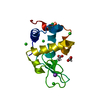 8cvuC  8cvvC  8cvwC  8cw1C  8cw3C  8cw5C  8cw6C  8cw7C  8cw8C  8cwbC  8cwcC  8cwdC 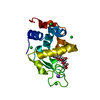 8cweC  8cwfC  8cwgC  8cwhC 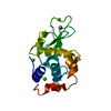 1ieeS S: Starting model for refinement C: citing same article ( |
|---|---|
| Similar structure data | |
| Experimental dataset #1 | Data reference: 10.11577/1873469 / Data set type: diffraction image data |
-Links
 PDBj
PDBj


- Assembly
| Deposited unit | 
| ||||||||||
|---|---|---|---|---|---|---|---|---|---|---|---|
| 1 |
| ||||||||||
| Unit cell |
| ||||||||||
| Components on special symmetry positions |
|
-Components
| #1: Protein | Mass: 14331.160 Da / Num. of mol.: 1 / Fragment: lyzozyme / Source method: obtained synthetically / Source: (synth.) | ||||||||
|---|---|---|---|---|---|---|---|---|---|
| #2: Chemical | ChemComp-NA /   Mass: 22.990 Da / Num. of mol.: 1 / Source method: obtained synthetically / Formula: Na Mass: 22.990 Da / Num. of mol.: 1 / Source method: obtained synthetically / Formula: Na | ||||||||
| #3: Chemical |   Mass: 59.044 Da / Num. of mol.: 2 / Source method: obtained synthetically / Formula: C2H3O2 Mass: 59.044 Da / Num. of mol.: 2 / Source method: obtained synthetically / Formula: C2H3O2#4: Chemical | ChemComp-CL /   Mass: 35.453 Da / Num. of mol.: 5 / Source method: isolated from a natural source / Formula: Cl Mass: 35.453 Da / Num. of mol.: 5 / Source method: isolated from a natural source / Formula: Cl#5: Water | ChemComp-HOH / |  Mass: 18.015 Da / Num. of mol.: 90 / Source method: isolated from a natural source / Formula: H2O Mass: 18.015 Da / Num. of mol.: 90 / Source method: isolated from a natural source / Formula: H2OHas ligand of interest | N | Has protein modification | Y | |
-Experimental details
-Experiment
| Experiment | Method: X-RAY DIFFRACTION / Number of used crystals: 1 |
|---|
- Sample preparation
| Crystal | Density Matthews: 2.11 Å3/Da / Density % sol: 41.65 % |
|---|---|
| Crystal grow | Temperature: 291 K / Method: batch mode / pH: 3 Details: Lysozyme [20 mg/ml dissolved in 0.1 M sodium acetate at pH 3.0] mixed with precipitant [28% (w/v) NaCl, 8% (w/v) PEG6000 and 0.1 M sodium acetate at pH 3.0] in a 1:1 ratio |
-Data collection
| Diffraction | Mean temperature: 291 K / Serial crystal experiment: Y |
|---|---|
| Diffraction source | Source: FREE ELECTRON LASER / Site: SACLA  / Beamline: BL3 / Wavelength: 1.24 Å / Beamline: BL3 / Wavelength: 1.24 Å |
| Detector | Type: MPCCD / Detector: CCD / Date: Nov 29, 2017 |
| Radiation | Protocol: SINGLE WAVELENGTH / Monochromatic (M) / Laue (L): M / Scattering type: x-ray |
| Radiation wavelength | Wavelength: 1.24 Å / Relative weight: 1 |
| Reflection | Resolution: 1.57→28.11 Å / Num. obs: 2208287 / % possible obs: 99.97 % / Redundancy: 125.46 % / Biso Wilson estimate: 15.49 Å2 / CC1/2: 0.992 / Net I/σ(I): 10.844 |
| Reflection shell | Resolution: 1.57→1.6 Å / Num. unique obs: 56144 / CC1/2: 0.445 |
| Serial crystallography sample delivery | Method: injection |
- Processing
| Software |
| |||||||||||||||||||||||||||||||||||||||||||||||||
|---|---|---|---|---|---|---|---|---|---|---|---|---|---|---|---|---|---|---|---|---|---|---|---|---|---|---|---|---|---|---|---|---|---|---|---|---|---|---|---|---|---|---|---|---|---|---|---|---|---|---|
| Refinement | Method to determine structure: MOLECULAR REPLACEMENT Starting model: 1iee Resolution: 1.57→28.11 Å / SU ML: 0.1561 / Cross valid method: FREE R-VALUE / σ(F): 1.99 / Phase error: 15.4445 Stereochemistry target values: GeoStd + Monomer Library + CDL v1.2
| |||||||||||||||||||||||||||||||||||||||||||||||||
| Solvent computation | Shrinkage radii: 0.9 Å / VDW probe radii: 1.11 Å / Solvent model: FLAT BULK SOLVENT MODEL | |||||||||||||||||||||||||||||||||||||||||||||||||
| Displacement parameters | Biso mean: 20.29 Å2 | |||||||||||||||||||||||||||||||||||||||||||||||||
| Refinement step | Cycle: LAST / Resolution: 1.57→28.11 Å
| |||||||||||||||||||||||||||||||||||||||||||||||||
| Refine LS restraints |
| |||||||||||||||||||||||||||||||||||||||||||||||||
| LS refinement shell |
|
