 Movie
Movie Controller

[English] 日本語
 Yorodumi
- PDB-8cu8: Cryo-EM structure of Ferritin 2 from Caenorhabditis elegans, FTN-2 -
+ Open data

- Basic information
| Entry | Database: PDB / ID: 8cu8 | ||||||
|---|---|---|---|---|---|---|---|
| Title | Cryo-EM structure of Ferritin 2 from Caenorhabditis elegans, FTN-2 | ||||||
 Components Components | Ferritin | ||||||
 Keywords Keywords | OXIDOREDUCTASE / ferroxidase | ||||||
| Function / homology |  Function and homology information Function and homology informationIron uptake and transport / Neutrophil degranulation / ferroxidase / ferroxidase activity / ferric iron binding / iron ion transport / ferrous iron binding / intracellular iron ion homeostasis / defense response to Gram-positive bacterium / identical protein binding / cytoplasm Similarity search - Function | ||||||
| Biological species |  | ||||||
| Method | ELECTRON MICROSCOPY / single particle reconstruction / cryo EM / Resolution: 1.91 Å | ||||||
 Authors Authors | Malcolm, T.R. / Brown, H.G. / Hanssen, E. | ||||||
| Funding support |  Australia, 1items Australia, 1items
| ||||||
 Citation Citation | Journal: To Be Published Title: Biochemical Characterization of Caenorhabditis elegans Ferritins Authors: Mubarak, S.M.M. / Malcolm, T.R. / Brown, H.G. / Hanssen, E. / Maher, M.J. / McColl, G. / Jameson, G.N.L. | ||||||
| History |
|
- Structure visualization
| Structure viewer | Molecule: MolmilJmol/JSmol |
|---|
- Downloads & links
-Download
| PDBx/mmCIF format | 8cu8.cif.gz | 736.8 KB | Display | PDBx/mmCIF format |
|---|---|---|---|---|
| PDB format | pdb8cu8.ent.gz | 617.5 KB | Display | PDB format |
| PDBx/mmJSON format | 8cu8.json.gz | Tree view | PDBx/mmJSON format | |
| Others |  Other downloads Other downloads |
-Validation report
| Arichive directory | https://data.pdbj.org/pub/pdb/validation_reports/cu/8cu8ftp://data.pdbj.org/pub/pdb/validation_reports/cu/8cu8 | HTTPS FTP |
|---|
-Related structure data
| Related structure data |  26996MC M: map data used to model this data C: citing same article ( |
|---|---|
| Similar structure data |
-Links
 PDBj
PDBj


- Assembly
| Deposited unit | 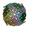
|
|---|---|
| 1 |
|
-Components
| #1: Protein | Mass: 19371.547 Da / Num. of mol.: 24 Source method: isolated from a genetically manipulated source Source: (gene. exp.)  #2: Chemical | ChemComp-FE /   Mass: 55.845 Da / Num. of mol.: 24 / Source method: obtained synthetically / Formula: Fe Mass: 55.845 Da / Num. of mol.: 24 / Source method: obtained synthetically / Formula: Fe#3: Water | ChemComp-HOH / |  Mass: 18.015 Da / Num. of mol.: 2271 / Source method: isolated from a natural source / Formula: H2O Mass: 18.015 Da / Num. of mol.: 2271 / Source method: isolated from a natural source / Formula: H2OHas ligand of interest | N | |
|---|
-Experimental details
-Experiment
| Experiment | Method: ELECTRON MICROSCOPY |
|---|---|
| EM experiment | Aggregation state: PARTICLE / 3D reconstruction method: single particle reconstruction |
- Sample preparation
| Component | Name: Homo 24-mer of Ferritin 2 / Type: COMPLEX / Entity ID: #1 / Source: RECOMBINANT |
|---|---|
| Source (natural) | Organism: |
| Source (recombinant) | Organism: |
| Buffer solution | pH: 8 |
| Specimen | Embedding applied: NO / Shadowing applied: NO / Staining applied: NO / Vitrification applied: YES |
| Specimen support | Grid material: GOLD / Grid type: UltrAuFoil R1.2/1.3 |
| Vitrification | Cryogen name: ETHANE |
- Electron microscopy imaging
| Experimental equipment |  Model: Titan Krios / Image courtesy: FEI Company |
|---|---|
| Microscopy | Model: TFS KRIOS |
| Electron gun | Electron source:  FIELD EMISSION GUN / Accelerating voltage: 300 kV / Illumination mode: FLOOD BEAM FIELD EMISSION GUN / Accelerating voltage: 300 kV / Illumination mode: FLOOD BEAM |
| Electron lens | Mode: BRIGHT FIELD / Nominal defocus max: 1400 nm / Nominal defocus min: 600 nm |
| Image recording | Electron dose: 47.6 e/Å2 / Film or detector model: FEI FALCON IV (4k x 4k) |
- Processing
| CTF correction | Type: PHASE FLIPPING AND AMPLITUDE CORRECTION |
|---|---|
| 3D reconstruction | Resolution: 1.91 Å / Resolution method: FSC 0.143 CUT-OFF / Num. of particles: 398769 / Symmetry type: POINT |
