 Movie
Movie Controller

[English] 日本語
 Yorodumi
- PDB-8ctq: Crystal structure of engineered phospholipase D mutant superPLD 2-48 -
+ Open data

- Basic information
| Entry | Database: PDB / ID: 8ctq | ||||||||||||||||||
|---|---|---|---|---|---|---|---|---|---|---|---|---|---|---|---|---|---|---|---|
| Title | Crystal structure of engineered phospholipase D mutant superPLD 2-48 | ||||||||||||||||||
 Components Components | Phospholipase D | ||||||||||||||||||
 Keywords Keywords | HYDROLASE / Enzyme / phospholipase / PLD / phospholipid | ||||||||||||||||||
| Function / homology |  Function and homology information Function and homology informationphosphatidyltransferase activity / cardiolipin biosynthetic process / phospholipase D / phospholipase D activity Similarity search - Function | ||||||||||||||||||
| Biological species |  Streptomyces sp. PMF (bacteria) Streptomyces sp. PMF (bacteria) | ||||||||||||||||||
| Method |  X-RAY DIFFRACTION / SYNCHROTRON / MOLECULAR REPLACEMENT / Resolution: 1.85 Å X-RAY DIFFRACTION / SYNCHROTRON / MOLECULAR REPLACEMENT / Resolution: 1.85 Å | ||||||||||||||||||
 Authors Authors | Tei, R. / Bagde, S.R. / Fromme, J.C. / Baskin, J.M. | ||||||||||||||||||
| Funding support |  United States, 5items United States, 5items
| ||||||||||||||||||
 Citation Citation | Journal: Nat.Chem. / Year: 2023 Title: Activity-based directed evolution of a membrane editor in mammalian cells. Authors: Tei, R. / Bagde, S.R. / Fromme, J.C. / Baskin, J.M. | ||||||||||||||||||
| History |
|
- Structure visualization
| Structure viewer | Molecule: MolmilJmol/JSmol |
|---|
- Downloads & links
-Download
| PDBx/mmCIF format | 8ctq.cif.gz | 231 KB | Display | PDBx/mmCIF format |
|---|---|---|---|---|
| PDB format | pdb8ctq.ent.gz | 148.4 KB | Display | PDB format |
| PDBx/mmJSON format | 8ctq.json.gz | Tree view | PDBx/mmJSON format | |
| Others |  Other downloads Other downloads |
-Validation report
| Summary document | 8ctq_validation.pdf.gz | 934.2 KB | Display | wwPDB validaton report |
|---|---|---|---|---|
| Full document | 8ctq_full_validation.pdf.gz | 938.1 KB | Display | |
| Data in XML | 8ctq_validation.xml.gz | 21.2 KB | Display | |
| Data in CIF | 8ctq_validation.cif.gz | 31.1 KB | Display | |
| Arichive directory | https://data.pdbj.org/pub/pdb/validation_reports/ct/8ctqftp://data.pdbj.org/pub/pdb/validation_reports/ct/8ctq | HTTPS FTP |
-Related structure data
| Related structure data |  8ctpC  1v0yS C: citing same article ( S: Starting model for refinement |
|---|---|
| Similar structure data |
-Links
 PDBj
PDBj- Assembly
| Deposited unit | 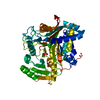
| ||||||||||||
|---|---|---|---|---|---|---|---|---|---|---|---|---|---|
| 1 |
| ||||||||||||
| Unit cell |
|
-Components
| #1: Protein | Mass: 54796.168 Da / Num. of mol.: 1 Mutation: K57R, A59V, K109R, P245A, V264I, G328S, G381V, G406S, G429D Source method: isolated from a genetically manipulated source Source: (gene. exp.) Streptomyces sp. PMF (bacteria) / Strain: PMF / Production host: |
|---|---|
| #2: Chemical | ChemComp-PO4 /   Mass: 94.971 Da / Num. of mol.: 1 / Source method: obtained synthetically / Formula: PO4 / Feature type: SUBJECT OF INVESTIGATION Mass: 94.971 Da / Num. of mol.: 1 / Source method: obtained synthetically / Formula: PO4 / Feature type: SUBJECT OF INVESTIGATION |
| #3: Water | ChemComp-HOH /  Mass: 18.015 Da / Num. of mol.: 284 / Source method: isolated from a natural source / Formula: H2O Mass: 18.015 Da / Num. of mol.: 284 / Source method: isolated from a natural source / Formula: H2O |
| Has ligand of interest | Y |
| Has protein modification | Y |
-Experimental details
-Experiment
| Experiment | Method: X-RAY DIFFRACTION / Number of used crystals: 1 |
|---|
- Sample preparation
| Crystal | Density Matthews: 2.01 Å3/Da / Density % sol: 39 % |
|---|---|
| Crystal grow | Temperature: 291 K / Method: vapor diffusion, sitting drop / pH: 4.4 Details: 21% PEG8000, 0.15 M lithium sulfate, citrate-NaOH, pH 4.4 |
-Data collection
| Diffraction | Mean temperature: 100 K / Serial crystal experiment: N |
|---|---|
| Diffraction source | Source: SYNCHROTRON / Site: APS / Beamline: 24-ID-E / Wavelength: 0.9792 Å |
| Detector | Type: DECTRIS EIGER X 16M / Detector: PIXEL / Date: Nov 17, 2021 |
| Radiation | Protocol: SINGLE WAVELENGTH / Monochromatic (M) / Laue (L): M / Scattering type: x-ray |
| Radiation wavelength | Wavelength: 0.9792 Å / Relative weight: 1 |
| Reflection | Resolution: 1.85→56.09 Å / Num. obs: 40025 / % possible obs: 99.15 % / Redundancy: 13.1 % / Biso Wilson estimate: 28.98 Å2 / CC1/2: 0.998 / Net I/σ(I): 13.55 |
| Reflection shell | Resolution: 1.85→1.918 Å / Mean I/σ(I) obs: 2.35 / Num. unique obs: 3612 / CC1/2: 0.879 |
- Processing
| Software |
| |||||||||||||||||||||||||||||||||||||||||||||||||||||||||||||||||||||||||||||||||||||||||||||||||||||||||
|---|---|---|---|---|---|---|---|---|---|---|---|---|---|---|---|---|---|---|---|---|---|---|---|---|---|---|---|---|---|---|---|---|---|---|---|---|---|---|---|---|---|---|---|---|---|---|---|---|---|---|---|---|---|---|---|---|---|---|---|---|---|---|---|---|---|---|---|---|---|---|---|---|---|---|---|---|---|---|---|---|---|---|---|---|---|---|---|---|---|---|---|---|---|---|---|---|---|---|---|---|---|---|---|---|---|---|
| Refinement | Method to determine structure: MOLECULAR REPLACEMENT Starting model: PDB entry 1V0Y Resolution: 1.85→56.09 Å / SU ML: 0.1925 / Cross valid method: FREE R-VALUE / σ(F): 1.36 / Phase error: 21.1422 Stereochemistry target values: GeoStd + Monomer Library + CDL v1.2
| |||||||||||||||||||||||||||||||||||||||||||||||||||||||||||||||||||||||||||||||||||||||||||||||||||||||||
| Solvent computation | Shrinkage radii: 0.9 Å / VDW probe radii: 1.1 Å / Solvent model: FLAT BULK SOLVENT MODEL | |||||||||||||||||||||||||||||||||||||||||||||||||||||||||||||||||||||||||||||||||||||||||||||||||||||||||
| Displacement parameters | Biso mean: 36.66 Å2 | |||||||||||||||||||||||||||||||||||||||||||||||||||||||||||||||||||||||||||||||||||||||||||||||||||||||||
| Refinement step | Cycle: LAST / Resolution: 1.85→56.09 Å
| |||||||||||||||||||||||||||||||||||||||||||||||||||||||||||||||||||||||||||||||||||||||||||||||||||||||||
| Refine LS restraints |
| |||||||||||||||||||||||||||||||||||||||||||||||||||||||||||||||||||||||||||||||||||||||||||||||||||||||||
| LS refinement shell |
|
