 Movie
Movie Controller

[English] 日本語
 Yorodumi
- PDB-8ct0: Crystal structure of FAD reductase CtcQ from Kitasatospora aureof... -
+ Open data

- Basic information
| Entry | Database: PDB / ID: 8ct0 | ||||||
|---|---|---|---|---|---|---|---|
| Title | Crystal structure of FAD reductase CtcQ from Kitasatospora aureofaciens in complex with FAD and NAD | ||||||
 Components Components | Flavin reductase (NADH) | ||||||
 Keywords Keywords | FLAVOPROTEIN / Reductase / Chlortetracycline / FAD binding / Biosysthesis / Natural product | ||||||
| Function / homology |  Function and homology information Function and homology informationflavin reductase (NADH) / flavin reductase (NADH) activity / FMN binding Similarity search - Function | ||||||
| Biological species |  Kitasatospora aureofaciens (bacteria) Kitasatospora aureofaciens (bacteria) | ||||||
| Method |  X-RAY DIFFRACTION / SYNCHROTRON / MOLECULAR REPLACEMENT / Resolution: 2.45 Å X-RAY DIFFRACTION / SYNCHROTRON / MOLECULAR REPLACEMENT / Resolution: 2.45 Å | ||||||
 Authors Authors | Hou, C. / Tsodikov, O.V. | ||||||
| Funding support | 1items
| ||||||
 Citation Citation | Journal: To Be Published Title: Crystal structures and complex formation of halogenase CtcP and FAD reductase CtcQ from the chlortetracycline biosynthetic pathway Authors: Hou, C. / Garneau-Tsodikova, S. / Tsodikov, O.V. | ||||||
| History |
|
- Structure visualization
| Structure viewer | Molecule: MolmilJmol/JSmol |
|---|
- Downloads & links
-Download
| PDBx/mmCIF format | 8ct0.cif.gz | 281.8 KB | Display | PDBx/mmCIF format |
|---|---|---|---|---|
| PDB format | pdb8ct0.ent.gz | 229.5 KB | Display | PDB format |
| PDBx/mmJSON format | 8ct0.json.gz | Tree view | PDBx/mmJSON format | |
| Others |  Other downloads Other downloads |
-Validation report
| Summary document | 8ct0_validation.pdf.gz | 9.1 MB | Display | wwPDB validaton report |
|---|---|---|---|---|
| Full document | 8ct0_full_validation.pdf.gz | 9.2 MB | Display | |
| Data in XML | 8ct0_validation.xml.gz | 55.6 KB | Display | |
| Data in CIF | 8ct0_validation.cif.gz | 73 KB | Display | |
| Arichive directory | https://data.pdbj.org/pub/pdb/validation_reports/ct/8ct0ftp://data.pdbj.org/pub/pdb/validation_reports/ct/8ct0 | HTTPS FTP |
-Related structure data
| Related structure data | 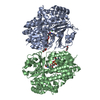 7v0bC 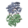 7v0dC  4l82S S: Starting model for refinement C: citing same article ( |
|---|---|
| Similar structure data |
-Links
 PDBj
PDBj

- Assembly
| Deposited unit | 
| ||||||||
|---|---|---|---|---|---|---|---|---|---|
| 1 | 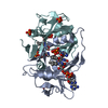
| ||||||||
| 2 | 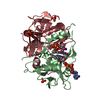
| ||||||||
| 3 | 
| ||||||||
| 4 | 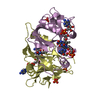
| ||||||||
| Unit cell |
|
-Components
-Protein , 1 types, 8 molecules ABCDEFGH
| #1: Protein | Mass: 20987.883 Da / Num. of mol.: 8 Source method: isolated from a genetically manipulated source Details: The C-terminal sequence LEHHHHHH is a C-terminal tag artifact. The residues are not present in the structure due to disorder. Source: (gene. exp.) Kitasatospora aureofaciens (bacteria) / Gene: ctcQ, B6264_18520, HS99_0013300 / Production host: |
|---|
-Non-polymers , 5 types, 263 molecules 


| #2: Chemical | ChemComp-FAD /  Mass: 785.550 Da / Num. of mol.: 14 / Source method: obtained synthetically / Formula: C27H33N9O15P2 / Feature type: SUBJECT OF INVESTIGATION / Comment: FAD*YM Mass: 785.550 Da / Num. of mol.: 14 / Source method: obtained synthetically / Formula: C27H33N9O15P2 / Feature type: SUBJECT OF INVESTIGATION / Comment: FAD*YM#3: Chemical | ChemComp-PO4 /  Mass: 94.971 Da / Num. of mol.: 12 / Source method: obtained synthetically / Formula: PO4 / Feature type: SUBJECT OF INVESTIGATION Mass: 94.971 Da / Num. of mol.: 12 / Source method: obtained synthetically / Formula: PO4 / Feature type: SUBJECT OF INVESTIGATION#4: Chemical | ChemComp-NAD / |  Mass: 663.425 Da / Num. of mol.: 1 / Source method: obtained synthetically / Formula: C21H27N7O14P2 / Feature type: SUBJECT OF INVESTIGATION / Comment: NAD*YM Mass: 663.425 Da / Num. of mol.: 1 / Source method: obtained synthetically / Formula: C21H27N7O14P2 / Feature type: SUBJECT OF INVESTIGATION / Comment: NAD*YM#5: Chemical | ChemComp-NAI / |  Mass: 665.441 Da / Num. of mol.: 1 / Source method: obtained synthetically / Formula: C21H29N7O14P2 Mass: 665.441 Da / Num. of mol.: 1 / Source method: obtained synthetically / Formula: C21H29N7O14P2#6: Water | ChemComp-HOH / | Mass: 18.015 Da / Num. of mol.: 235 / Source method: isolated from a natural source / Formula: H2O |
|---|
-Details
| Has ligand of interest | Y |
|---|
-Experimental details
-Experiment
| Experiment | Method: X-RAY DIFFRACTION / Number of used crystals: 1 |
|---|
- Sample preparation
| Crystal | Density Matthews: 2.12 Å3/Da / Density % sol: 41.92 % |
|---|---|
| Crystal grow | Temperature: 294 K / Method: vapor diffusion, hanging drop / pH: 7.5 / Details: 0.6 M KH2PO4,0.6 M NaH2PO4, 0.1 M Tris pH 7.5 |
-Data collection
| Diffraction | Mean temperature: 100 K / Serial crystal experiment: N |
|---|---|
| Diffraction source | Source: SYNCHROTRON / Site: APS  / Beamline: 22-ID / Wavelength: 1 Å / Beamline: 22-ID / Wavelength: 1 Å |
| Detector | Type: DECTRIS EIGER X 16M / Detector: PIXEL / Date: Feb 11, 2020 |
| Radiation | Protocol: SINGLE WAVELENGTH / Monochromatic (M) / Laue (L): M / Scattering type: x-ray |
| Radiation wavelength | Wavelength: 1 Å / Relative weight: 1 |
| Reflection | Resolution: 2.45→50 Å / Num. obs: 49072 / % possible obs: 96.4 % / Observed criterion σ(I): 2 / Redundancy: 3.4 % / CC1/2: 0.978 / Rmerge(I) obs: 0.138 / Net I/σ(I): 8.9 |
| Reflection shell | Resolution: 2.45→2.49 Å / Redundancy: 3.2 % / Rmerge(I) obs: 0.568 / Num. unique obs: 2399 / CC1/2: 0.696 / % possible all: 95.4 |
- Processing
| Software |
| ||||||||||||||||||||||||||||||||||||||||||||||||||||||||||||
|---|---|---|---|---|---|---|---|---|---|---|---|---|---|---|---|---|---|---|---|---|---|---|---|---|---|---|---|---|---|---|---|---|---|---|---|---|---|---|---|---|---|---|---|---|---|---|---|---|---|---|---|---|---|---|---|---|---|---|---|---|---|
| Refinement | Method to determine structure: MOLECULAR REPLACEMENT Starting model: 4L82 Resolution: 2.45→34.97 Å / Cor.coef. Fo:Fc: 0.932 / Cor.coef. Fo:Fc free: 0.877 / Cross valid method: THROUGHOUT / σ(F): 0 / ESU R: 1.112 / ESU R Free: 0.316 / Stereochemistry target values: MAXIMUM LIKELIHOOD Details: HYDROGENS HAVE BEEN ADDED IN THE RIDING POSITIONS U VALUES : REFINED INDIVIDUALLY
| ||||||||||||||||||||||||||||||||||||||||||||||||||||||||||||
| Solvent computation | Ion probe radii: 0.8 Å / Shrinkage radii: 0.8 Å / VDW probe radii: 1.2 Å / Solvent model: MASK | ||||||||||||||||||||||||||||||||||||||||||||||||||||||||||||
| Displacement parameters | Biso max: 69.59 Å2 / Biso mean: 21.327 Å2 / Biso min: 2.94 Å2
| ||||||||||||||||||||||||||||||||||||||||||||||||||||||||||||
| Refinement step | Cycle: final / Resolution: 2.45→34.97 Å
| ||||||||||||||||||||||||||||||||||||||||||||||||||||||||||||
| Refine LS restraints |
| ||||||||||||||||||||||||||||||||||||||||||||||||||||||||||||
| LS refinement shell | Resolution: 2.45→2.513 Å / Rfactor Rfree error: 0 / Total num. of bins used: 20
|
