 Movie
Movie Controller

[English] 日本語
 Yorodumi
- PDB-8cp7: Structure of the disulfide-locked substrate binding protein HiSiaP. -
+ Open data

- Basic information
| Entry | Database: PDB / ID: 8cp7 | ||||||
|---|---|---|---|---|---|---|---|
| Title | Structure of the disulfide-locked substrate binding protein HiSiaP. | ||||||
 Components Components | Sialic acid-binding periplasmic protein SiaP | ||||||
 Keywords Keywords | SUGAR BINDING PROTEIN / Substrate binding protein / TRAP / sialic acid | ||||||
| Function / homology | TRAP transporter solute receptor, DctP family / TRAP transporter solute receptor DctP / TRAP transporter solute receptor DctP superfamily / Bacterial extracellular solute-binding protein, family 7 / transmembrane transport / outer membrane-bounded periplasmic space / N-acetyl-beta-neuraminic acid / Sialic acid-binding periplasmic protein SiaP Function and homology information Function and homology information | ||||||
| Biological species |  Haemophilus influenzae (bacteria) Haemophilus influenzae (bacteria) | ||||||
| Method |  X-RAY DIFFRACTION / SYNCHROTRON / MOLECULAR REPLACEMENT / Resolution: 1.9 Å X-RAY DIFFRACTION / SYNCHROTRON / MOLECULAR REPLACEMENT / Resolution: 1.9 Å | ||||||
 Authors Authors | Kim, Y. / Peter, M.F. / Hagelueken, G. | ||||||
| Funding support |  Germany, 1items Germany, 1items
| ||||||
 Citation Citation | Journal: Nat Commun / Year: 2024 Title: Conformational coupling of the sialic acid TRAP transporter HiSiaQM with its substrate binding protein HiSiaP. Authors: Peter, M.F. / Ruland, J.A. / Kim, Y. / Hendricks, P. / Schneberger, N. / Siebrasse, J.P. / Thomas, G.H. / Kubitscheck, U. / Hagelueken, G. | ||||||
| History |
|
- Structure visualization
| Structure viewer | Molecule: MolmilJmol/JSmol |
|---|
- Downloads & links
-Download
| PDBx/mmCIF format | 8cp7.cif.gz | 96.1 KB | Display | PDBx/mmCIF format |
|---|---|---|---|---|
| PDB format | pdb8cp7.ent.gz | 57.4 KB | Display | PDB format |
| PDBx/mmJSON format | 8cp7.json.gz | Tree view | PDBx/mmJSON format | |
| Others |  Other downloads Other downloads |
-Validation report
| Arichive directory | https://data.pdbj.org/pub/pdb/validation_reports/cp/8cp7ftp://data.pdbj.org/pub/pdb/validation_reports/cp/8cp7 | HTTPS FTP |
|---|
-Related structure data
| Similar structure data |
|---|
-Links
 PDBj
PDBj- Assembly
| Deposited unit | 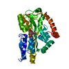
| ||||||||||||
|---|---|---|---|---|---|---|---|---|---|---|---|---|---|
| 1 |
| ||||||||||||
| Unit cell |
| ||||||||||||
| Components on special symmetry positions |
|
-Components
| #1: Protein | Mass: 34695.355 Da / Num. of mol.: 1 Source method: isolated from a genetically manipulated source Source: (gene. exp.) Haemophilus influenzae (bacteria) / Gene: siaP, HI_0146 / Production host: | ||||||
|---|---|---|---|---|---|---|---|
| #2: Sugar | ChemComp-SLB / 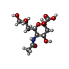  Type: D-saccharide, beta linking / Mass: 309.270 Da / Num. of mol.: 1 / Source method: obtained synthetically / Formula: C11H19NO9 / Feature type: SUBJECT OF INVESTIGATION Type: D-saccharide, beta linking / Mass: 309.270 Da / Num. of mol.: 1 / Source method: obtained synthetically / Formula: C11H19NO9 / Feature type: SUBJECT OF INVESTIGATION | ||||||
| #3: Chemical |   Mass: 65.409 Da / Num. of mol.: 2 / Source method: obtained synthetically / Formula: Zn Mass: 65.409 Da / Num. of mol.: 2 / Source method: obtained synthetically / Formula: Zn#4: Water | ChemComp-HOH / |  Mass: 18.015 Da / Num. of mol.: 134 / Source method: isolated from a natural source / Formula: H2O Mass: 18.015 Da / Num. of mol.: 134 / Source method: isolated from a natural source / Formula: H2OHas ligand of interest | Y | Has protein modification | Y | |
-Experimental details
-Experiment
| Experiment | Method: X-RAY DIFFRACTION / Number of used crystals: 1 |
|---|
- Sample preparation
| Crystal | Density Matthews: 2.45 Å3/Da / Density % sol: 49.84 % |
|---|---|
| Crystal grow | Temperature: 293 K / Method: vapor diffusion, sitting drop Details: 0.01 M Zinc chloride, 0.1 M Sodium acetate, pH 5.0, 20% w/v PEG 6000 |
-Data collection
| Diffraction | Mean temperature: 100 K / Serial crystal experiment: N |
|---|---|
| Diffraction source | Source: SYNCHROTRON / Site: PETRA III, EMBL c/o DESY / Beamline: P13 (MX1) / Wavelength: 0.826554 Å |
| Detector | Type: DECTRIS EIGER X 16M / Detector: PIXEL / Date: Sep 30, 2022 |
| Radiation | Protocol: SINGLE WAVELENGTH / Monochromatic (M) / Laue (L): M / Scattering type: x-ray |
| Radiation wavelength | Wavelength: 0.826554 Å / Relative weight: 1 |
| Reflection | Resolution: 1.9→42.04 Å / Num. obs: 26990 / % possible obs: 99.02 % / Redundancy: 2 % / Biso Wilson estimate: 33.96 Å2 / CC1/2: 1 / Rmerge(I) obs: 0.0289 / Net I/σ(I): 14.84 |
| Reflection shell | Resolution: 1.9→1.97 Å / Rmerge(I) obs: 0.613 / Num. unique obs: 2437 / CC1/2: 0.721 |
- Processing
| Software |
| ||||||||||||||||||||||||||||||||||||||||||||||||||||||||||||||||||||||||||||||||||||
|---|---|---|---|---|---|---|---|---|---|---|---|---|---|---|---|---|---|---|---|---|---|---|---|---|---|---|---|---|---|---|---|---|---|---|---|---|---|---|---|---|---|---|---|---|---|---|---|---|---|---|---|---|---|---|---|---|---|---|---|---|---|---|---|---|---|---|---|---|---|---|---|---|---|---|---|---|---|---|---|---|---|---|---|---|---|
| Refinement | Method to determine structure: MOLECULAR REPLACEMENT / Resolution: 1.9→42.04 Å / SU ML: 0.2686 / Cross valid method: FREE R-VALUE / σ(F): 1.34 / Phase error: 30.5777 Stereochemistry target values: GeoStd + Monomer Library + CDL v1.2
| ||||||||||||||||||||||||||||||||||||||||||||||||||||||||||||||||||||||||||||||||||||
| Solvent computation | Shrinkage radii: 0.9 Å / VDW probe radii: 1.11 Å / Solvent model: FLAT BULK SOLVENT MODEL | ||||||||||||||||||||||||||||||||||||||||||||||||||||||||||||||||||||||||||||||||||||
| Displacement parameters | Biso mean: 39.37 Å2 | ||||||||||||||||||||||||||||||||||||||||||||||||||||||||||||||||||||||||||||||||||||
| Refinement step | Cycle: LAST / Resolution: 1.9→42.04 Å
| ||||||||||||||||||||||||||||||||||||||||||||||||||||||||||||||||||||||||||||||||||||
| Refine LS restraints |
| ||||||||||||||||||||||||||||||||||||||||||||||||||||||||||||||||||||||||||||||||||||
| LS refinement shell |
|
