[English] 日本語
Yorodumi
- PDB-8cnc: Structure of compound 1 bound KMT9 -

+
Open data


ID or keywords:

Loading...

-
Basic information

Entry
Database: PDB / ID: 8cnc
TitleStructure of compound 1 bound KMT9
Components
  • Methyltransferase N6AMT1
  • Multifunctional methyltransferase subunit TRM112-like protein
KeywordsTRANSFERASE / protein methyltransferase / inhibitor / SAM analogue
Function / homology
Function and homology information


arsonoacetate metabolic process / methylarsonite methyltransferase activity / histone H4K12 methyltransferase activity / eRF1 methyltransferase complex / protein-glutamine N-methyltransferase activity / peptidyl-glutamine methylation / rRNA (guanine-N7)-methylation / tRNA methyltransferase activator activity / toxin metabolic process / site-specific DNA-methyltransferase (adenine-specific) activity ...arsonoacetate metabolic process / methylarsonite methyltransferase activity / histone H4K12 methyltransferase activity / eRF1 methyltransferase complex / protein-glutamine N-methyltransferase activity / peptidyl-glutamine methylation / rRNA (guanine-N7)-methylation / tRNA methyltransferase activator activity / toxin metabolic process / site-specific DNA-methyltransferase (adenine-specific) activity / tRNA modification in the nucleus and cytosol / Methylation / protein methyltransferase activity / tRNA methylation / positive regulation of rRNA processing / S-adenosylmethionine-dependent methyltransferase activity / S-adenosyl-L-methionine binding / rRNA modification in the nucleus and cytosol / rRNA methylation / Eukaryotic Translation Termination / transcription initiation-coupled chromatin remodeling / Transferases; Transferring one-carbon groups; Methyltransferases / methylation / positive regulation of cell growth / nucleic acid binding / protein heterodimerization activity / perinuclear region of cytoplasm / protein-containing complex / nucleoplasm / nucleus / cytosol
Similarity search - Function
Eukaryotic/archaeal PrmC-related / Multifunctional methyltransferase subunit Trm112 / Trm112-like / Trm112p-like protein / Methyltransferase small domain / Methyltransferase small domain / N-6 Adenine-specific DNA methylases signature. / DNA methylase, N-6 adenine-specific, conserved site / S-adenosyl-L-methionine-dependent methyltransferase superfamily
Similarity search - Domain/homology
Chem-6D6 / Multifunctional methyltransferase subunit TRM112-like protein / Methyltransferase N6AMT1
Similarity search - Component
Biological speciesHomo sapiens (human)
MethodX-RAY DIFFRACTION / SYNCHROTRON / MOLECULAR REPLACEMENT / Resolution: 1.46 Å
AuthorsSheng, W.
Funding support Germany, 6items
OrganizationGrant numberCountry
German Research Foundation (DFG)SFB850 Germany
German Research Foundation (DFG)SFB992 Germany
German Research Foundation (DFG)SFB1381 Germany
German Research Foundation (DFG)Schu688/15-1 Germany
German Research Foundation (DFG)FR01-374 Germany
German Research Foundation (DFG)EXC-2189 Germany
CitationJournal: To Be Published
Title: Structure of compound 1 bound KMT9
Authors: Sheng, W. / Eric, M. / Roland, S.
History
DepositionFeb 22, 2023Deposition site: PDBE / Processing site: PDBE
Revision 1.0Mar 6, 2024Provider: repository / Type: Initial release

-
Structure visualization

Structure viewerMolecule:
MolmilJmol/JSmol

Downloads & links

-
Assembly

Deposited unit
A: Methyltransferase N6AMT1
B: Multifunctional methyltransferase subunit TRM112-like protein
hetero molecules


Theoretical massNumber of molelcules
Total (without water)36,5013
Polymers36,0762
Non-polymers4241
Water3,099172
1


  • Idetical with deposited unit
  • defined by author&software
  • Evidence: gel filtration
TypeNameSymmetry operationNumber
identity operation1_555x,y,z1
Buried area2380 Å2
ΔGint-12 kcal/mol
Surface area13770 Å2
MethodPISA
Unit cell
Length a, b, c (Å)110.344, 110.344, 129.875
Angle α, β, γ (deg.)90.00, 90.00, 120.00
Int Tables number178
Space group name H-MP6122
Components on special symmetry positions
IDModelComponents
11A-489-

HOH

-
Components

#1: Protein Methyltransferase N6AMT1 / HemK methyltransferase family member 2 / M.HsaHemK2P / Lysine N-methyltransferase 9 / ...HemK methyltransferase family member 2 / M.HsaHemK2P / Lysine N-methyltransferase 9 / Methylarsonite methyltransferase N6AMT1 / Protein N(5)-glutamine methyltransferase


Mass: 21804.064 Da / Num. of mol.: 1
Source method: isolated from a genetically manipulated source
Source: (gene. exp.) Homo sapiens (human) / Gene: N6AMT1, C21orf127, HEMK2, KMT9, PRED28 / Production host: Escherichia coli (E. coli)
References: UniProt: Q9Y5N5, Transferases; Transferring one-carbon groups; Methyltransferases
#2: Protein Multifunctional methyltransferase subunit TRM112-like protein / tRNA methyltransferase 112 homolog


Mass: 14272.412 Da / Num. of mol.: 1
Source method: isolated from a genetically manipulated source
Source: (gene. exp.) Homo sapiens (human) / Gene: TRMT112, AD-001, HSPC152, HSPC170 / Production host: Escherichia coli (E. coli) / References: UniProt: Q9UI30
#3: Chemical ChemComp-6D6 / 5'-{[(3S)-3-amino-3-carboxypropyl](3-aminopropyl)amino}-5'-deoxyadenosine


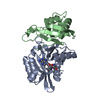
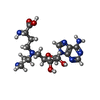
Mass: 424.455 Da / Num. of mol.: 1 / Source method: obtained synthetically / Formula: C17H28N8O5
#4: Water ChemComp-HOH / water / Water


Mass: 18.015 Da / Num. of mol.: 172 / Source method: isolated from a natural source / Formula: H2O
Has ligand of interestN

-
Experimental details

-
Experiment

ExperimentMethod: X-RAY DIFFRACTION / Number of used crystals: 1

-
Sample preparation

CrystalDensity Matthews: 3.16 Å3/Da / Density % sol: 61.12 %
Crystal growTemperature: 293 K / Method: vapor diffusion, sitting drop / pH: 8 / Details: 1.3 M Na3Citrate, 0.1M Tris ph 8.0

-
Data collection

DiffractionMean temperature: 100 K / Serial crystal experiment: N
Diffraction sourceSource: SYNCHROTRON / Site: SLS / Beamline: X06DA / Wavelength: 1 Å
DetectorType: DECTRIS PILATUS 2M-F / Detector: PIXEL / Date: Feb 11, 2019
RadiationProtocol: SINGLE WAVELENGTH / Monochromatic (M) / Laue (L): M / Scattering type: x-ray
Radiation wavelengthWavelength: 1 Å / Relative weight: 1
ReflectionResolution: 1.46→47.78 Å / Num. obs: 80857 / % possible obs: 99.9 % / Redundancy: 38.9 % / CC1/2: 1 / Net I/σ(I): 1.6
Reflection shellResolution: 1.46→8 Å / Num. unique obs: 3867 / CC1/2: 0.579

-
Processing

Software
NameVersionClassification
REFMAC5refinement
Aimlessdata scaling
XDSdata reduction
PHASERphasing
RefinementMethod to determine structure: MOLECULAR REPLACEMENT / Resolution: 1.46→47.83 Å / Cor.coef. Fo:Fc: 0.973 / Cor.coef. Fo:Fc free: 0.96 / SU B: 2.011 / SU ML: 0.034 / Cross valid method: THROUGHOUT / ESU R: 0.051 / ESU R Free: 0.051 / Stereochemistry target values: MAXIMUM LIKELIHOOD / Details: HYDROGENS HAVE BEEN ADDED IN THE RIDING POSITIONS
RfactorNum. reflection% reflectionSelection details
Rfree0.1833 4070 5 %RANDOM
Rwork0.15179 ---
obs0.15337 76713 99.94 %-
Solvent computationIon probe radii: 0.8 Å / Shrinkage radii: 0.8 Å / VDW probe radii: 1.2 Å / Solvent model: MASK
Displacement parametersBiso mean: 19.019 Å2
Baniso -1Baniso -2Baniso -3
1-0 Å20 Å20 Å2
2--0 Å2-0 Å2
3----0 Å2
Refinement stepCycle: 1 / Resolution: 1.46→47.83 Å
ProteinNucleic acidLigandSolventTotal
Num. atoms2401 0 30 172 2603
Refine LS restraints
Refine-IDTypeDev idealDev ideal targetNumber
X-RAY DIFFRACTIONr_bond_refined_d0.0130.0122555
X-RAY DIFFRACTIONr_bond_other_d0.0010.0162407
X-RAY DIFFRACTIONr_angle_refined_deg1.7621.6363489
X-RAY DIFFRACTIONr_angle_other_deg0.611.5455629
X-RAY DIFFRACTIONr_dihedral_angle_1_deg6.3375330
X-RAY DIFFRACTIONr_dihedral_angle_2_deg10.7691016
X-RAY DIFFRACTIONr_dihedral_angle_3_deg14.62410434
X-RAY DIFFRACTIONr_dihedral_angle_4_deg
X-RAY DIFFRACTIONr_chiral_restr0.0970.2406
X-RAY DIFFRACTIONr_gen_planes_refined0.010.022917
X-RAY DIFFRACTIONr_gen_planes_other0.0010.02466
X-RAY DIFFRACTIONr_nbd_refined
X-RAY DIFFRACTIONr_nbd_other
X-RAY DIFFRACTIONr_nbtor_refined
X-RAY DIFFRACTIONr_nbtor_other
X-RAY DIFFRACTIONr_xyhbond_nbd_refined
X-RAY DIFFRACTIONr_xyhbond_nbd_other
X-RAY DIFFRACTIONr_metal_ion_refined
X-RAY DIFFRACTIONr_metal_ion_other
X-RAY DIFFRACTIONr_symmetry_vdw_refined
X-RAY DIFFRACTIONr_symmetry_vdw_other
X-RAY DIFFRACTIONr_symmetry_hbond_refined
X-RAY DIFFRACTIONr_symmetry_hbond_other
X-RAY DIFFRACTIONr_symmetry_metal_ion_refined
X-RAY DIFFRACTIONr_symmetry_metal_ion_other
X-RAY DIFFRACTIONr_mcbond_it1.9551.131290
X-RAY DIFFRACTIONr_mcbond_other1.9511.1291290
X-RAY DIFFRACTIONr_mcangle_it2.2051.6981624
X-RAY DIFFRACTIONr_mcangle_other2.2051.6991625
X-RAY DIFFRACTIONr_scbond_it3.31.4351265
X-RAY DIFFRACTIONr_scbond_other3.2981.4351266
X-RAY DIFFRACTIONr_scangle_it
X-RAY DIFFRACTIONr_scangle_other3.522.0481864
X-RAY DIFFRACTIONr_long_range_B_refined3.22316.1032674
X-RAY DIFFRACTIONr_long_range_B_other3.22316.1032675
X-RAY DIFFRACTIONr_rigid_bond_restr5.55934962
X-RAY DIFFRACTIONr_sphericity_free
X-RAY DIFFRACTIONr_sphericity_bonded
LS refinement shellResolution: 1.461→1.499 Å / Total num. of bins used: 20
RfactorNum. reflection% reflection
Rfree0.267 285 -
Rwork0.235 5562 -
obs--99.44 %
Refinement TLS params.

Method: refined / Refine-ID: X-RAY DIFFRACTION

IDL112)L122)L132)L222)L232)L332)S11 (Å °)S12 (Å °)S13 (Å °)S21 (Å °)S22 (Å °)S23 (Å °)S31 (Å °)S32 (Å °)S33 (Å °)T112)T122)T132)T222)T232)T332)Origin x (Å)Origin y (Å)Origin z (Å)
10.63870.1489-0.04322.0508-0.44931.8512-0.02920.07790.0776-0.12950.00510.0609-0.06770.05650.02410.038-0.0031-0.01910.01190.00910.0161-42.4518-6.9587-16.506
22.36320.3419-0.49841.3149-0.01422.0041-0.0007-0.04560.0128-0.0017-0.05340.06520.0567-0.11130.05410.0319-0.0002-0.00720.0104-0.00690.0073-47.1569-26.7944-2.4665
Refinement TLS group
IDRefine-IDRefine TLS-IDAuth asym-IDAuth seq-ID
1X-RAY DIFFRACTION1A20 - 213
2X-RAY DIFFRACTION2B2 - 119

+
About Yorodumi

-
News

-
Feb 9, 2022. New format data for meta-information of EMDB entries

New format data for meta-information of EMDB entries

  • Version 3 of the EMDB header file is now the official format.
  • The previous official version 1.9 will be removed from the archive.

Related info.:EMDB header

External links:wwPDB to switch to version 3 of the EMDB data model

-
Aug 12, 2020. Covid-19 info

Covid-19 info

URL: https://pdbj.org/emnavi/covid19.php

New page: Covid-19 featured information page in EM Navigator.

Related info.:Covid-19 info / Mar 5, 2020. Novel coronavirus structure data

+
Mar 5, 2020. Novel coronavirus structure data

Novel coronavirus structure data

Related info.:Yorodumi Speices / Aug 12, 2020. Covid-19 info

External links:COVID-19 featured content - PDBj / Molecule of the Month (242):Coronavirus Proteases

+
Jan 31, 2019. EMDB accession codes are about to change! (news from PDBe EMDB page)

EMDB accession codes are about to change! (news from PDBe EMDB page)

  • The allocation of 4 digits for EMDB accession codes will soon come to an end. Whilst these codes will remain in use, new EMDB accession codes will include an additional digit and will expand incrementally as the available range of codes is exhausted. The current 4-digit format prefixed with “EMD-” (i.e. EMD-XXXX) will advance to a 5-digit format (i.e. EMD-XXXXX), and so on. It is currently estimated that the 4-digit codes will be depleted around Spring 2019, at which point the 5-digit format will come into force.
  • The EM Navigator/Yorodumi systems omit the EMD- prefix.

Related info.:Q: What is EMD? / ID/Accession-code notation in Yorodumi/EM Navigator

External links:EMDB Accession Codes are Changing Soon! / Contact to PDBj

+
Jul 12, 2017. Major update of PDB

Major update of PDB

  • wwPDB released updated PDB data conforming to the new PDBx/mmCIF dictionary.
  • This is a major update changing the version number from 4 to 5, and with Remediation, in which all the entries are updated.
  • In this update, many items about electron microscopy experimental information are reorganized (e.g. em_software).
  • Now, EM Navigator and Yorodumi are based on the updated data.

External links:wwPDB Remediation / Enriched Model Files Conforming to OneDep Data Standards Now Available in the PDB FTP Archive

-
Yorodumi

Thousand views of thousand structures

  • Yorodumi is a browser for structure data from EMDB, PDB, SASBDB, etc.
  • This page is also the successor to EM Navigator detail page, and also detail information page/front-end page for Omokage search.
  • The word "yorodu" (or yorozu) is an old Japanese word meaning "ten thousand". "mi" (miru) is to see.

Related info.:EMDB / PDB / SASBDB / Comparison of 3 databanks / Yorodumi Search / Aug 31, 2016. New EM Navigator & Yorodumi / Yorodumi Papers / Jmol/JSmol / Function and homology information / Changes in new EM Navigator and Yorodumi

Read more