 Movie
Movie Controller

[English] 日本語
 Yorodumi
- PDB-8cmx: Structure of sphingosine-1-phosphate lyase (SPL) from Aspergillus... -
+ Open data

- Basic information
| Entry | Database: PDB / ID: 8cmx | ||||||||||||
|---|---|---|---|---|---|---|---|---|---|---|---|---|---|
| Title | Structure of sphingosine-1-phosphate lyase (SPL) from Aspergillus fumigatus | ||||||||||||
 Components Components | Sphinganine-1-phosphate aldolase BST1, putative | ||||||||||||
 Keywords Keywords | LYASE / sphingosine / sphingosine-1-phosphate / S1P | ||||||||||||
| Function / homology |  Function and homology information Function and homology informationsphinganine-1-phosphate aldolase / sphinganine-1-phosphate aldolase activity / perinuclear endoplasmic reticulum / cortical endoplasmic reticulum / carboxylic acid metabolic process / sphingolipid catabolic process / cellular response to starvation / calcium-mediated signaling / pyridoxal phosphate binding / endoplasmic reticulum / identical protein binding Similarity search - Function | ||||||||||||
| Biological species |  | ||||||||||||
| Method |  X-RAY DIFFRACTION / SYNCHROTRON / MOLECULAR REPLACEMENT / Resolution: 3.46 Å X-RAY DIFFRACTION / SYNCHROTRON / MOLECULAR REPLACEMENT / Resolution: 3.46 Å | ||||||||||||
 Authors Authors | Catalano, F. / Pampalone, G. | ||||||||||||
| Funding support | 3items
| ||||||||||||
 Citation Citation | Journal: Sci Rep / Year: 2023 Title: Dual species sphingosine-1-phosphate lyase inhibitors to combine antifungal and anti-inflammatory activities in cystic fibrosis: a feasibility study. Authors: Cellini, B. / Pampalone, G. / Camaioni, E. / Pariano, M. / Catalano, F. / Zelante, T. / Dindo, M. / Macchioni, L. / Di Veroli, A. / Galarini, R. / Paoletti, F. / Davidescu, M. / Stincardini, ...Authors: Cellini, B. / Pampalone, G. / Camaioni, E. / Pariano, M. / Catalano, F. / Zelante, T. / Dindo, M. / Macchioni, L. / Di Veroli, A. / Galarini, R. / Paoletti, F. / Davidescu, M. / Stincardini, C. / Vascelli, G. / Bellet, M.M. / Saba, J. / Giovagnoli, S. / Giardina, G. / Romani, L. / Costantini, C. | ||||||||||||
| History |
|
- Structure visualization
| Structure viewer | Molecule: MolmilJmol/JSmol |
|---|
- Downloads & links
-Download
| PDBx/mmCIF format | 8cmx.cif.gz | 196.4 KB | Display | PDBx/mmCIF format |
|---|---|---|---|---|
| PDB format | pdb8cmx.ent.gz | 154.2 KB | Display | PDB format |
| PDBx/mmJSON format | 8cmx.json.gz | Tree view | PDBx/mmJSON format | |
| Others |  Other downloads Other downloads |
-Validation report
| Summary document | 8cmx_validation.pdf.gz | 452.9 KB | Display | wwPDB validaton report |
|---|---|---|---|---|
| Full document | 8cmx_full_validation.pdf.gz | 473.9 KB | Display | |
| Data in XML | 8cmx_validation.xml.gz | 36.4 KB | Display | |
| Data in CIF | 8cmx_validation.cif.gz | 48.6 KB | Display | |
| Arichive directory | https://data.pdbj.org/pub/pdb/validation_reports/cm/8cmxftp://data.pdbj.org/pub/pdb/validation_reports/cm/8cmx | HTTPS FTP |
-Related structure data

-Links
 PDBj
PDBj- Assembly
| Deposited unit | 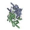
| |||||||||
|---|---|---|---|---|---|---|---|---|---|---|
| 1 |
| |||||||||
| Unit cell |
| |||||||||
| Noncrystallographic symmetry (NCS) | NCS domain:
|
-Components
| #1: Protein | Mass: 54601.070 Da / Num. of mol.: 2 Source method: isolated from a genetically manipulated source Details: Missing 81 residues at N-terminal / Source: (gene. exp.)  References: UniProt: Q4WPU3, sphinganine-1-phosphate aldolase Has ligand of interest | N | |
|---|
-Experimental details
-Experiment
| Experiment | Method: X-RAY DIFFRACTION / Number of used crystals: 1 |
|---|
- Sample preparation
| Crystal | Density Matthews: 2.63 Å3/Da / Density % sol: 53.34 % / Description: hexagonal yellow crystals |
|---|---|
| Crystal grow | Temperature: 294 K / Method: vapor diffusion, hanging drop / pH: 7.5 Details: 0.1M Hepes sodium pH 7.5 1.4 M Sodium citrate tribasic dihydrate DMSO 1% Cryoprotection = mother liquor + 20% Glycerol AfuSPL = 93 microM |
-Data collection
| Diffraction | Mean temperature: 100 K / Serial crystal experiment: N |
|---|---|
| Diffraction source | Source: SYNCHROTRON / Site: ELETTRA  / Beamline: 11.2C / Wavelength: 1 Å / Beamline: 11.2C / Wavelength: 1 Å |
| Detector | Type: DECTRIS PILATUS 6M / Detector: PIXEL / Date: Sep 21, 2022 |
| Radiation | Protocol: SINGLE WAVELENGTH / Monochromatic (M) / Laue (L): M / Scattering type: x-ray |
| Radiation wavelength | Wavelength: 1 Å / Relative weight: 1 |
| Reflection | Resolution: 3.46→101.5 Å / Num. obs: 12600 / % possible obs: 95.1 % / Redundancy: 36.7 % / CC1/2: 0.998 / Rmerge(I) obs: 0.317 / Net I/σ(I): 15.1 |
| Reflection shell | Resolution: 3.46→3.74 Å / Redundancy: 39 % / Mean I/σ(I) obs: 1.5 / Num. unique obs: 631 / CC1/2: 0.572 / % possible all: 71.9 |
- Processing
| Software |
| ||||||||||||||||||||||||||||||||||||||||||||||||||||||||||||||||||||||||||||||||||||||||||||||||||||||||||||||||||||||||||||||||||||||||||||||||||||||||||||||||||||||||||||||||||||||
|---|---|---|---|---|---|---|---|---|---|---|---|---|---|---|---|---|---|---|---|---|---|---|---|---|---|---|---|---|---|---|---|---|---|---|---|---|---|---|---|---|---|---|---|---|---|---|---|---|---|---|---|---|---|---|---|---|---|---|---|---|---|---|---|---|---|---|---|---|---|---|---|---|---|---|---|---|---|---|---|---|---|---|---|---|---|---|---|---|---|---|---|---|---|---|---|---|---|---|---|---|---|---|---|---|---|---|---|---|---|---|---|---|---|---|---|---|---|---|---|---|---|---|---|---|---|---|---|---|---|---|---|---|---|---|---|---|---|---|---|---|---|---|---|---|---|---|---|---|---|---|---|---|---|---|---|---|---|---|---|---|---|---|---|---|---|---|---|---|---|---|---|---|---|---|---|---|---|---|---|---|---|---|---|
| Refinement | Method to determine structure: MOLECULAR REPLACEMENT / Resolution: 3.46→100.02 Å / Cor.coef. Fo:Fc: 0.891 / Cor.coef. Fo:Fc free: 0.897 / SU B: 51.092 / SU ML: 0.724 / Cross valid method: THROUGHOUT / ESU R Free: 0.918 / Stereochemistry target values: MAXIMUM LIKELIHOOD / Details: HYDROGENS HAVE BEEN ADDED IN THE RIDING POSITIONS
| ||||||||||||||||||||||||||||||||||||||||||||||||||||||||||||||||||||||||||||||||||||||||||||||||||||||||||||||||||||||||||||||||||||||||||||||||||||||||||||||||||||||||||||||||||||||
| Solvent computation | Ion probe radii: 0.8 Å / Shrinkage radii: 0.8 Å / VDW probe radii: 1.2 Å / Solvent model: MASK | ||||||||||||||||||||||||||||||||||||||||||||||||||||||||||||||||||||||||||||||||||||||||||||||||||||||||||||||||||||||||||||||||||||||||||||||||||||||||||||||||||||||||||||||||||||||
| Displacement parameters | Biso mean: 121.81 Å2
| ||||||||||||||||||||||||||||||||||||||||||||||||||||||||||||||||||||||||||||||||||||||||||||||||||||||||||||||||||||||||||||||||||||||||||||||||||||||||||||||||||||||||||||||||||||||
| Refinement step | Cycle: 1 / Resolution: 3.46→100.02 Å
| ||||||||||||||||||||||||||||||||||||||||||||||||||||||||||||||||||||||||||||||||||||||||||||||||||||||||||||||||||||||||||||||||||||||||||||||||||||||||||||||||||||||||||||||||||||||
| Refine LS restraints |
|