 Movie
Movie Controller

[English] 日本語
 Yorodumi
- PDB-8cm8: Galectin-8 N-terminal carbohydrate recognition domain in complex ... -
+ Open data

- Basic information
| Entry | Database: PDB / ID: 8cm8 | ||||||
|---|---|---|---|---|---|---|---|
| Title | Galectin-8 N-terminal carbohydrate recognition domain in complex with 4-(bromophenyl)phthalazinone D-galactal ligand | ||||||
 Components Components | Galectin-8 | ||||||
 Keywords Keywords | SUGAR BINDING PROTEIN / Galectin / phthalazinone / inhibitor | ||||||
| Function / homology |  Function and homology information Function and homology informationlymphatic endothelial cell migration / xenophagy / cellular response to virus / integrin binding / carbohydrate binding / cytoplasmic vesicle / extracellular space / membrane / cytosol / cytoplasm Similarity search - Function | ||||||
| Biological species |  Homo sapiens (human) Homo sapiens (human) | ||||||
| Method |  X-RAY DIFFRACTION / SYNCHROTRON / MOLECULAR REPLACEMENT / Resolution: 1.22 Å X-RAY DIFFRACTION / SYNCHROTRON / MOLECULAR REPLACEMENT / Resolution: 1.22 Å | ||||||
 Authors Authors | Van Klaveren, S. / Hakansson, M. / Diehl, C. / Nilsson, N.J. | ||||||
| Funding support | European Union, 1items
| ||||||
 Citation Citation | Journal: Acs Med.Chem.Lett. / Year: 2024 Title: Galectin-8N-Selective 4-Halophenylphthalazinone-Galactals Double pi-Stack in a Unique Pocket. Authors: van Klaveren, S. / Hassan, M. / Hakansson, M. / Johnsson, R.E. / Larsson, J. / Jakopin, Z. / Anderluh, M. / Leffler, H. / Tomasic, T. / Nilsson, U.J. | ||||||
| History |
|
- Structure visualization
| Structure viewer | Molecule: MolmilJmol/JSmol |
|---|
- Downloads & links
-Download
| PDBx/mmCIF format | 8cm8.cif.gz | 351.5 KB | Display | PDBx/mmCIF format |
|---|---|---|---|---|
| PDB format | pdb8cm8.ent.gz | 218 KB | Display | PDB format |
| PDBx/mmJSON format | 8cm8.json.gz | Tree view | PDBx/mmJSON format | |
| Others |  Other downloads Other downloads |
-Validation report
| Arichive directory | https://data.pdbj.org/pub/pdb/validation_reports/cm/8cm8ftp://data.pdbj.org/pub/pdb/validation_reports/cm/8cm8 | HTTPS FTP |
|---|
-Related structure data
| Similar structure data |
|---|
-Links
 PDBj
PDBj- Assembly
| Deposited unit | 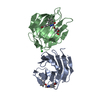
| ||||||||
|---|---|---|---|---|---|---|---|---|---|
| 1 |
| ||||||||
| Unit cell |
|
-Components
| #1: Protein | Mass: 35854.055 Da / Num. of mol.: 2 Source method: isolated from a genetically manipulated source Source: (gene. exp.) Homo sapiens (human) / Gene: LGALS8 / Production host:  #2: Chemical | Mass: 459.290 Da / Num. of mol.: 2 / Source method: obtained synthetically / Formula: C21H19BrN2O5 / Feature type: SUBJECT OF INVESTIGATION #3: Chemical | ChemComp-CL / |   Mass: 35.453 Da / Num. of mol.: 1 / Source method: obtained synthetically / Formula: Cl Mass: 35.453 Da / Num. of mol.: 1 / Source method: obtained synthetically / Formula: Cl#4: Water | ChemComp-HOH / |  Mass: 18.015 Da / Num. of mol.: 289 / Source method: isolated from a natural source / Formula: H2O Mass: 18.015 Da / Num. of mol.: 289 / Source method: isolated from a natural source / Formula: H2OHas ligand of interest | Y | Has protein modification | N | |
|---|
-Experimental details
-Experiment
| Experiment | Method: X-RAY DIFFRACTION / Number of used crystals: 1 |
|---|
- Sample preparation
| Crystal | Density Matthews: 2.17 Å3/Da / Density % sol: 43.38 % |
|---|---|
| Crystal grow | Temperature: 293.5 K / Method: vapor diffusion, hanging drop / pH: 8 Details: A co-crystal with lactose, grown from 12 mg/ml galectin-8N in 10 mM lactose, 10 mM Tris/HCl pH 8.0, 1 mM TCEP and 150 mM sodium chloride mixed with 25% (w/v) PEG 2000 MME) in a hanging drop, ...Details: A co-crystal with lactose, grown from 12 mg/ml galectin-8N in 10 mM lactose, 10 mM Tris/HCl pH 8.0, 1 mM TCEP and 150 mM sodium chloride mixed with 25% (w/v) PEG 2000 MME) in a hanging drop, was used to soak the compound by transferring crystals in three steps to soaking drops. These were 2 uL drops with a soaking solution of 20% ethylene glycol, 25% PEG 2000 MME, 10 mM Tris pH 8, 1 mM TCEP, 50 mM NaCl, and 2 mM compound 10. The incubation times in the first two drops were about 10 min and in the third drop about 24 h. Then the crystals were transferred to a cryo solution with the same constituents and flash-frozen in liquid nitrogen. |
-Data collection
| Diffraction | Mean temperature: 100 K / Serial crystal experiment: N |
|---|---|
| Diffraction source | Source: SYNCHROTRON / Site: Diamond  / Beamline: I24 / Wavelength: 0.9999 Å / Beamline: I24 / Wavelength: 0.9999 Å |
| Detector | Type: DECTRIS PILATUS3 X 6M / Detector: PIXEL / Date: Sep 18, 2021 Details: Oxford Danfysik/SESO Two stage demagnification using two K-B pairs of bimorph type mirrors |
| Radiation | Monochromator: ACCEL Fixed exit Double Crystal / Protocol: SINGLE WAVELENGTH / Monochromatic (M) / Laue (L): M / Scattering type: x-ray |
| Radiation wavelength | Wavelength: 0.9999 Å / Relative weight: 1 |
| Reflection | Resolution: 1.22→50.19 Å / Num. obs: 82202 / % possible obs: 99.9 % / Redundancy: 11.9 % / Rmerge(I) obs: 0.076 / Rpim(I) all: 0.023 / Rrim(I) all: 0.079 / Χ2: 0.99 / Net I/σ(I): 13.5 |
| Reflection shell | Resolution: 1.22→1.252 Å / Redundancy: 9 % / Rmerge(I) obs: 0.05 / Num. unique obs: 6317 / Rpim(I) all: 0.022 / Rrim(I) all: 0.054 / % possible all: 99.9 |
- Processing
| Software |
| |||||||||||||||||||||||||||||||||||||||||||||||||||||||||||||||||||||||||||||||||||||||||||||||||||||||||||||||||||||||||||||||||||||||||||||||||||||||||||
|---|---|---|---|---|---|---|---|---|---|---|---|---|---|---|---|---|---|---|---|---|---|---|---|---|---|---|---|---|---|---|---|---|---|---|---|---|---|---|---|---|---|---|---|---|---|---|---|---|---|---|---|---|---|---|---|---|---|---|---|---|---|---|---|---|---|---|---|---|---|---|---|---|---|---|---|---|---|---|---|---|---|---|---|---|---|---|---|---|---|---|---|---|---|---|---|---|---|---|---|---|---|---|---|---|---|---|---|---|---|---|---|---|---|---|---|---|---|---|---|---|---|---|---|---|---|---|---|---|---|---|---|---|---|---|---|---|---|---|---|---|---|---|---|---|---|---|---|---|---|---|---|---|---|---|---|---|
| Refinement | Method to determine structure: MOLECULAR REPLACEMENT / Resolution: 1.22→50.186 Å / Cor.coef. Fo:Fc: 0.975 / Cor.coef. Fo:Fc free: 0.964 / SU B: 2.242 / SU ML: 0.042 / Cross valid method: THROUGHOUT / ESU R: 0.047 / ESU R Free: 0.049 Details: Hydrogens have been added in their riding positions
| |||||||||||||||||||||||||||||||||||||||||||||||||||||||||||||||||||||||||||||||||||||||||||||||||||||||||||||||||||||||||||||||||||||||||||||||||||||||||||
| Solvent computation | Ion probe radii: 0.8 Å / Shrinkage radii: 0.8 Å / VDW probe radii: 1.2 Å / Solvent model: MASK BULK SOLVENT | |||||||||||||||||||||||||||||||||||||||||||||||||||||||||||||||||||||||||||||||||||||||||||||||||||||||||||||||||||||||||||||||||||||||||||||||||||||||||||
| Displacement parameters | Biso mean: 21.302 Å2
| |||||||||||||||||||||||||||||||||||||||||||||||||||||||||||||||||||||||||||||||||||||||||||||||||||||||||||||||||||||||||||||||||||||||||||||||||||||||||||
| Refinement step | Cycle: LAST / Resolution: 1.22→50.186 Å
| |||||||||||||||||||||||||||||||||||||||||||||||||||||||||||||||||||||||||||||||||||||||||||||||||||||||||||||||||||||||||||||||||||||||||||||||||||||||||||
| Refine LS restraints |
| |||||||||||||||||||||||||||||||||||||||||||||||||||||||||||||||||||||||||||||||||||||||||||||||||||||||||||||||||||||||||||||||||||||||||||||||||||||||||||
| LS refinement shell |
|
