 ムービー
ムービー コントローラー


+ データを開く

- 基本情報
基本情報
| 登録情報 | データベース: PDB / ID: 8cka | |||||||||
|---|---|---|---|---|---|---|---|---|---|---|
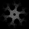
| タイトル | Deinococcus radidurans HPI S-layer | |||||||||
 要素 要素 | Hexagonally packed intermediate-layer surface protein | |||||||||
 キーワード キーワード | STRUCTURAL PROTEIN / HPI S-layer | |||||||||
| 機能・相同性 | S-layer / cell wall organization / Prokaryotic membrane lipoprotein lipid attachment site profile. / extracellular region / Hexagonally packed intermediate-layer surface protein 機能・相同性情報 機能・相同性情報 | |||||||||
| 生物種 |  Deinococcus radiodurans (放射線耐性) Deinococcus radiodurans (放射線耐性) | |||||||||
| 手法 | 電子顕微鏡法 / 単粒子再構成法 / クライオ電子顕微鏡法 / 解像度: 2.52 Å | |||||||||
 データ登録者 データ登録者 | von Kuegelgen, A. / Yamashita, K. / Murshudov, G. / Bharat, T. | |||||||||
| 資金援助 |  英国, 2件 英国, 2件
| |||||||||
 引用 引用 | ジャーナル: Proc Natl Acad Sci U S A / 年: 2023 タイトル: Interdigitated immunoglobulin arrays form the hyperstable surface layer of the extremophilic bacterium . 著者: Andriko von Kügelgen / Sofie van Dorst / Keitaro Yamashita / Danielle L Sexton / Elitza I Tocheva / Garib Murshudov / Vikram Alva / Tanmay A M Bharat /   要旨: is an atypical diderm bacterium with a remarkable ability to tolerate various environmental stresses, due in part to its complex cell envelope encapsulated within a hyperstable surface layer (S- ... is an atypical diderm bacterium with a remarkable ability to tolerate various environmental stresses, due in part to its complex cell envelope encapsulated within a hyperstable surface layer (S-layer). Despite decades of research on this cell envelope, atomic structural details of the S-layer have remained obscure. In this study, we report the electron cryomicroscopy structure of the S-layer, showing how it is formed by the Hexagonally Packed Intermediate-layer (HPI) protein arranged in a planar hexagonal lattice. The HPI protein forms an array of immunoglobulin-like folds within the S-layer, with each monomer extending into the adjacent hexamer, resulting in a highly interconnected, stable, sheet-like arrangement. Using electron cryotomography and subtomogram averaging from focused ion beam-milled cells, we have obtained a structure of the cellular S-layer, showing how this HPI S-layer coats native membranes on the surface of cells. Our S-layer structure from the diderm bacterium shows similarities to immunoglobulin-like domain-containing S-layers from monoderm bacteria and archaea, highlighting common features in cell surface organization across different domains of life, with connotations on the evolution of immunoglobulin-based molecular recognition systems in eukaryotes. | |||||||||
| 履歴 |
|
- 構造の表示
構造の表示
| 構造ビューア | 分子: MolmilJmol/JSmol |
|---|
- ダウンロードとリンク
-ダウンロード
| PDBx/mmCIF形式 | 8cka.cif.gz | 197.7 KB | 表示 | PDBx/mmCIF形式 |
|---|---|---|---|---|
| PDB形式 | pdb8cka.ent.gz | 144.9 KB | 表示 | PDB形式 |
| PDBx/mmJSON形式 | 8cka.json.gz | ツリー表示 | PDBx/mmJSON形式 | |
| その他 |  その他のダウンロード その他のダウンロード |
-検証レポート
| 文書・要旨 | 8cka_validation.pdf.gz | 1.6 MB | 表示 | wwPDB検証レポート |
|---|---|---|---|---|
| 文書・詳細版 | 8cka_full_validation.pdf.gz | 1.6 MB | 表示 | |
| XML形式データ | 8cka_validation.xml.gz | 45.9 KB | 表示 | |
| CIF形式データ | 8cka_validation.cif.gz | 68 KB | 表示 | |
| アーカイブディレクトリ | https://data.pdbj.org/pub/pdb/validation_reports/ck/8ckaftp://data.pdbj.org/pub/pdb/validation_reports/ck/8cka | HTTPS FTP |
-関連構造データ
-リンク
 PDBj
PDBj


- 集合体
集合体
| 登録構造単位 | 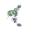
| ||||||||||||||||||||||||||||
|---|---|---|---|---|---|---|---|---|---|---|---|---|---|---|---|---|---|---|---|---|---|---|---|---|---|---|---|---|---|
| 1 | x 6
| ||||||||||||||||||||||||||||
| 非結晶学的対称性 (NCS) | NCS oper:
|
-要素
| #1: タンパク質 | 分子量: 99424.000 Da / 分子数: 2 / 由来タイプ: 天然 由来: (天然) Deinococcus radiodurans (strain ATCC 13939 / DSM 20539 / JCM 16871 / LMG 4051 / NBRC 15346 / NCIMB 9279 / R1 / VKM B-1422) (放射線耐性)参照: UniProt: P56867 Has protein modification | Y | |
|---|
-実験情報
-実験
| 実験 | 手法: 電子顕微鏡法 |
|---|---|
| EM実験 | 試料の集合状態: 2D ARRAY / 3次元再構成法: 単粒子再構成法 |
- 試料調製
試料調製
| 構成要素 | 名称: Structure of Hexagonally Packed Intermediate-layer (HPI) protein タイプ: ORGANELLE OR CELLULAR COMPONENT 詳細: Structure of Hexagonally Packed Intermediate-layer (HPI) protein Entity ID: all / 由来: NATURAL | |||||||||||||||||||||||||
|---|---|---|---|---|---|---|---|---|---|---|---|---|---|---|---|---|---|---|---|---|---|---|---|---|---|---|
| 分子量 | 実験値: NO | |||||||||||||||||||||||||
| 由来(天然) | 生物種: Deinococcus radiodurans (放射線耐性) / 細胞内の位置: extracellular | |||||||||||||||||||||||||
| 緩衝液 | pH: 7.5 詳細: 50 mM HEPES/NaOH pH=7.5, 150 mM NaCl, 5 mM MgCl2, 1 mM CaCl2 | |||||||||||||||||||||||||
| 緩衝液成分 |
| |||||||||||||||||||||||||
| 試料 | 包埋: NO / シャドウイング: NO / 染色: NO / 凍結: YES 詳細: In vitro isolate Hexagonally Packed Intermediate-layer (HPI) layer | |||||||||||||||||||||||||
| 試料支持 | 詳細: 15 mA / グリッドの材料: COPPER/RHODIUM / グリッドのサイズ: 200 divisions/in. / グリッドのタイプ: Quantifoil R2/2 | |||||||||||||||||||||||||
| 急速凍結 | 装置: FEI VITROBOT MARK IV / 凍結剤: NITROGEN / 湿度: 100 % / 凍結前の試料温度: 283.15 K 詳細: absorption for 60 sec and blotted for 5 sec with blot force -10 |
- 電子顕微鏡撮影
| 実験機器 |  モデル: Titan Krios / 画像提供: FEI Company |
|---|---|
| 顕微鏡 | モデル: FEI TITAN KRIOS |
| 電子銃 | 電子線源:  FIELD EMISSION GUN / 加速電圧: 300 kV / 照射モード: FLOOD BEAM FIELD EMISSION GUN / 加速電圧: 300 kV / 照射モード: FLOOD BEAM |
| 電子レンズ | モード: BRIGHT FIELD / 倍率(公称値): 81000 X / 倍率(補正後): 81000 X / 最大 デフォーカス(公称値): 5000 nm / 最小 デフォーカス(公称値): 2000 nm / Calibrated defocus min: 2000 nm / 最大 デフォーカス(補正後): 5000 nm / Cs: 2.7 mm / C2レンズ絞り径: 70 µm / アライメント法: COMA FREE |
| 試料ホルダ | 凍結剤: NITROGEN 試料ホルダーモデル: FEI TITAN KRIOS AUTOGRID HOLDER 最高温度: 70 K / 最低温度: 70 K |
| 撮影 | 平均露光時間: 3.43 sec. / 電子線照射量: 53.245 e/Å2 フィルム・検出器のモデル: GATAN K3 BIOQUANTUM (6k x 4k) 撮影したグリッド数: 1 / 実像数: 1002 |
| 電子光学装置 | エネルギーフィルター名称: GIF Quantum LS / 色収差補正装置: not used / エネルギーフィルタースリット幅: 20 eV / 球面収差補正装置: not used |
| 画像スキャン | 横: 5760 / 縦: 4092 |
- 解析
解析
| ソフトウェア | 名称: REFMAC / バージョン: 5.8.0403 / 分類: 精密化 | ||||||||||||||||||||||||||||||||||||||||||||||||||||||||||||||||||||||||||||||||||||||||||||||||||||||||||
|---|---|---|---|---|---|---|---|---|---|---|---|---|---|---|---|---|---|---|---|---|---|---|---|---|---|---|---|---|---|---|---|---|---|---|---|---|---|---|---|---|---|---|---|---|---|---|---|---|---|---|---|---|---|---|---|---|---|---|---|---|---|---|---|---|---|---|---|---|---|---|---|---|---|---|---|---|---|---|---|---|---|---|---|---|---|---|---|---|---|---|---|---|---|---|---|---|---|---|---|---|---|---|---|---|---|---|---|
| EMソフトウェア |
| ||||||||||||||||||||||||||||||||||||||||||||||||||||||||||||||||||||||||||||||||||||||||||||||||||||||||||
| 画像処理 | 詳細: Movies collected at the scope were clustered into optics groups based on the XML meta-data of the data-collection software EPU (Thermo Fisher Scientific) using a k-means algorithm implemented ...詳細: Movies collected at the scope were clustered into optics groups based on the XML meta-data of the data-collection software EPU (Thermo Fisher Scientific) using a k-means algorithm implemented in EPU_group_AFIS (https://github.com/DustinMorado/EPU_group_AFIS). Imported movies were motion-corrected, dose-weighted, and Fourier cropped (2x) with MotionCor2 implemented in RELION-3.1. Contrast transfer functions (CTFs) of the resulting motion-corrected micrographs were estimated using CTFFIND4. | ||||||||||||||||||||||||||||||||||||||||||||||||||||||||||||||||||||||||||||||||||||||||||||||||||||||||||
| CTF補正 | 詳細: RELION refinement with in-built CTF correction. The function is similar to a Wiener filter, so amplitude correction included. タイプ: PHASE FLIPPING AND AMPLITUDE CORRECTION | ||||||||||||||||||||||||||||||||||||||||||||||||||||||||||||||||||||||||||||||||||||||||||||||||||||||||||
| 粒子像の選択 | 選択した粒子像数: 882866 詳細: Initially, side views of S-layer sheets were first manually picked along the edge of the lattice using the helical picking tab in RELION while setting the helical rise to 40 Angstrom. Top and ...詳細: Initially, side views of S-layer sheets were first manually picked along the edge of the lattice using the helical picking tab in RELION while setting the helical rise to 40 Angstrom. Top and tilted views were manually picked at the central hexameric axis. Manually picked particles were extracted in 4x downsampled 100 x 100 boxes and classified using reference-free 2D classification inside RELION3.1. Class averages centered at a hexameric axis were used to automatically pick particles inside RELION3.1. Automatically picked particles were extracted in 4x downsampled 100x100 pixel2 boxes and classified using reference-free 2D classification. Particle coordinates belonging to class averages centered at the hexameric axis were used to train TOPAZ in 5x downsampled micrographs with the neural network architecture conv127. For the final reconstruction, particles were picked using TOPAZ and the previously trained neural network above. Additionally, top, bottom, and side views were picked using the reference-based autopicker inside RELION3.1, which TOPAZ did not readily identify. Particles were extracted in 4x downsampled 100x100 pixel2 boxes and classified using reference-free 2D classification inside RELION3.1. Particles belonging to class averages centered at the hexameric axis were combined, and particles within 30 Angstrom were removed to prevent duplication after alignment. All resulting particles were then re-extracted in 4x downsampled 100x100 pixel2 boxes. | ||||||||||||||||||||||||||||||||||||||||||||||||||||||||||||||||||||||||||||||||||||||||||||||||||||||||||
| 対称性 | 点対称性: C6 (6回回転対称) | ||||||||||||||||||||||||||||||||||||||||||||||||||||||||||||||||||||||||||||||||||||||||||||||||||||||||||
| 3次元再構成 | 解像度: 2.52 Å / 解像度の算出法: FSC 0.143 CUT-OFF / 粒子像の数: 55345 / アルゴリズム: FOURIER SPACE 詳細: Per-particle defocus, anisotropy magnification, and higher-order aberrations were refined inside RELION3.1, followed by another round of focused 3D auto refinement and Bayesian particle ...詳細: Per-particle defocus, anisotropy magnification, and higher-order aberrations were refined inside RELION3.1, followed by another round of focused 3D auto refinement and Bayesian particle polishing. The final map was obtained from 55,345 particles and post-processed using a soft mask focused on the central hexamer, including the dimeric bridge, yielding a global resolution of 2.52 Angstrom according to the gold standard Fourier shell correlation criterion of 0.143. クラス平均像の数: 1 / 対称性のタイプ: POINT | ||||||||||||||||||||||||||||||||||||||||||||||||||||||||||||||||||||||||||||||||||||||||||||||||||||||||||
| 原子モデル構築 | B value: 19.48 / プロトコル: AB INITIO MODEL / 空間: RECIPROCAL / Target criteria: Best Fit | ||||||||||||||||||||||||||||||||||||||||||||||||||||||||||||||||||||||||||||||||||||||||||||||||||||||||||
| 精密化 | 解像度: 2.52→2.52 Å / Cor.coef. Fo:Fc: 0.924 / SU B: 7.928 / SU ML: 0.163 / ESU R: 0.07 立体化学のターゲット値: MAXIMUM LIKELIHOOD WITH PHASES 詳細: HYDROGENS HAVE BEEN ADDED IN THE RIDING POSITIONS
| ||||||||||||||||||||||||||||||||||||||||||||||||||||||||||||||||||||||||||||||||||||||||||||||||||||||||||
| 溶媒の処理 | 溶媒モデル: PARAMETERS FOR MASK CACLULATION | ||||||||||||||||||||||||||||||||||||||||||||||||||||||||||||||||||||||||||||||||||||||||||||||||||||||||||
| 原子変位パラメータ | Biso mean: 75.352 Å2 | ||||||||||||||||||||||||||||||||||||||||||||||||||||||||||||||||||||||||||||||||||||||||||||||||||||||||||
| 精密化ステップ | サイクル: 1 / 合計: 6795 | ||||||||||||||||||||||||||||||||||||||||||||||||||||||||||||||||||||||||||||||||||||||||||||||||||||||||||
| 拘束条件 |
|