[English] 日本語
Yorodumi
- PDB-8cih: Structure of FL CINP -

+
Open data


ID or keywords:

Loading...

-
Basic information

Entry
Database: PDB / ID: 8cih
TitleStructure of FL CINP
ComponentsCyclin-dependent kinase 2-interacting protein
KeywordsREPLICATION / alpha-helical structure / protein complex subunit / DNA replication
Function / homologyCyclin-dependent kinase 2-interacting protein / preribosome binding / ribosomal large subunit biogenesis / DNA replication / cell cycle / cell division / DNA repair / nucleus / Cyclin-dependent kinase 2-interacting protein
Function and homology information
Biological speciesHomo sapiens (human)
MethodX-RAY DIFFRACTION / SYNCHROTRON / MOLECULAR REPLACEMENT / Resolution: 2 Å
AuthorsFoglizzo, M. / Zeqiraj, E.
Funding support United Kingdom, 3items
OrganizationGrant numberCountry
Medical Research Council (MRC, United Kingdom)MR/T029471/1 United Kingdom
Wellcome Trust222531/Z/21/Z United Kingdom
Biotechnology and Biological Sciences Research Council (BBSRC)BB/L015056/1 United Kingdom
Citation
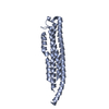
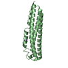
Journal: Cell / Year: 2024
Title: The SPATA5-SPATA5L1 ATPase complex directs replisome proteostasis to ensure genome integrity.
Authors: Vidhya Krishnamoorthy / Martina Foglizzo / Robert L Dilley / Angela Wu / Arindam Datta / Parul Dutta / Lisa J Campbell / Oksana Degtjarik / Laura J Musgrove / Antonio N Calabrese / Elton ...Authors: Vidhya Krishnamoorthy / Martina Foglizzo / Robert L Dilley / Angela Wu / Arindam Datta / Parul Dutta / Lisa J Campbell / Oksana Degtjarik / Laura J Musgrove / Antonio N Calabrese / Elton Zeqiraj / Roger A Greenberg /
Abstract: Ubiquitin-dependent unfolding of the CMG helicase by VCP/p97 is required to terminate DNA replication. Other replisome components are not processed in the same fashion, suggesting that additional ...Ubiquitin-dependent unfolding of the CMG helicase by VCP/p97 is required to terminate DNA replication. Other replisome components are not processed in the same fashion, suggesting that additional mechanisms underlie replication protein turnover. Here, we identify replisome factor interactions with a protein complex composed of AAA+ ATPases SPATA5-SPATA5L1 together with heterodimeric partners C1orf109-CINP (55LCC). An integrative structural biology approach revealed a molecular architecture of SPATA5-SPATA5L1 N-terminal domains interacting with C1orf109-CINP to form a funnel-like structure above a cylindrically shaped ATPase motor. Deficiency in the 55LCC complex elicited ubiquitin-independent proteotoxicity, replication stress, and severe chromosome instability. 55LCC showed ATPase activity that was specifically enhanced by replication fork DNA and was coupled to cysteine protease-dependent cleavage of replisome substrates in response to replication fork damage. These findings define 55LCC-mediated proteostasis as critical for replication fork progression and genome stability and provide a rationale for pathogenic variants seen in associated human neurodevelopmental disorders.
History
DepositionFeb 9, 2023Deposition site: PDBE / Processing site: PDBE
Revision 1.0Feb 21, 2024Provider: repository / Type: Initial release
Revision 1.1Mar 27, 2024Group: Database references / Category: citation / citation_author / Item: _citation.pdbx_database_id_DOI
Revision 1.2Apr 10, 2024Group: Database references / Category: citation / citation_author
Item: _citation.country / _citation.journal_abbrev ..._citation.country / _citation.journal_abbrev / _citation.journal_id_ASTM / _citation.journal_id_CSD / _citation.journal_id_ISSN / _citation.pdbx_database_id_PubMed / _citation.title / _citation_author.identifier_ORCID / _citation_author.name
Revision 1.3May 8, 2024Group: Database references / Category: citation / Item: _citation.journal_volume / _citation.page_first

-
Structure visualization

Structure viewerMolecule:
MolmilJmol/JSmol

Downloads & links

-
Assembly

Deposited unit
A: Cyclin-dependent kinase 2-interacting protein
B: Cyclin-dependent kinase 2-interacting protein
hetero molecules


Theoretical massNumber of molelcules
Total (without water)48,9993
Polymers48,9762
Non-polymers231
Water1,47782
1
A: Cyclin-dependent kinase 2-interacting protein


Theoretical massNumber of molelcules
Total (without water)24,4881
Polymers24,4881
Non-polymers00
Water181
TypeNameSymmetry operationNumber
identity operation1_555x,y,z1
2
B: Cyclin-dependent kinase 2-interacting protein
hetero molecules


Theoretical massNumber of molelcules
Total (without water)24,5112
Polymers24,4881
Non-polymers231
Water181
TypeNameSymmetry operationNumber
identity operation1_555x,y,z1
Unit cell
Length a, b, c (Å)91.722, 91.722, 134.368
Angle α, β, γ (deg.)90.00, 90.00, 120.00
Int Tables number169
Space group name H-MP61

-
Components

#1: Protein Cyclin-dependent kinase 2-interacting protein / CDK2-interacting protein


Mass: 24487.951 Da / Num. of mol.: 2
Source method: isolated from a genetically manipulated source
Source: (gene. exp.) Homo sapiens (human) / Gene: CINP / Production host: Escherichia coli (E. coli) / References: UniProt: Q9BW66
#2: Chemical ChemComp-NA / SODIUM ION


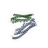
Mass: 22.990 Da / Num. of mol.: 1 / Source method: obtained synthetically / Formula: Na
#3: Water ChemComp-HOH / water / Water


Mass: 18.015 Da / Num. of mol.: 82 / Source method: isolated from a natural source / Formula: H2O
Has ligand of interestN

-
Experimental details

-
Experiment

ExperimentMethod: X-RAY DIFFRACTION / Number of used crystals: 1

-
Sample preparation

CrystalDensity Matthews: 3.97 Å3/Da / Density % sol: 69.01 %
Crystal growTemperature: 292.15 K / Method: vapor diffusion, sitting drop / pH: 6.2
Details: 25% (v/v) 1,2-propanediol, 10% (v/v) glycerol and 100 mM sodium potassium phosphate pH 6.2

-
Data collection

DiffractionMean temperature: 100 K / Serial crystal experiment: N
Diffraction sourceSource: SYNCHROTRON / Site: Diamond / Beamline: I04 / Wavelength: 0.9795 Å
DetectorType: DECTRIS EIGER2 XE 16M / Detector: PIXEL / Date: Nov 26, 2021
RadiationProtocol: SINGLE WAVELENGTH / Monochromatic (M) / Laue (L): M / Scattering type: x-ray
Radiation wavelengthWavelength: 0.9795 Å / Relative weight: 1
ReflectionResolution: 2→51.3 Å / Num. obs: 43390 / % possible obs: 95.7 % / Redundancy: 13.3 % / Biso Wilson estimate: 32.05 Å2 / CC1/2: 0.9 / Rmerge(I) obs: 0.122 / Rpim(I) all: 0.035 / Rrim(I) all: 0.127 / Net I/σ(I): 14.4
Reflection shellResolution: 2→2.1 Å / Num. unique obs: 40485 / CC1/2: 0.7 / % possible all: 93.8

-
Processing

Software
NameVersionClassification
PHENIX1.17.1_3660refinement
STARANISOdata scaling
AutoProcessdata scaling
PHASERphasing
autoPROCdata reduction
RefinementMethod to determine structure: MOLECULAR REPLACEMENT / Resolution: 2→51.3 Å / SU ML: 0.24 / Cross valid method: FREE R-VALUE / σ(F): 1.35 / Phase error: 24.7 / Stereochemistry target values: ML
RfactorNum. reflection% reflection
Rfree0.2313 2065 5.1 %
Rwork0.2104 --
obs0.2115 40484 93.77 %
Solvent computationShrinkage radii: 0.9 Å / VDW probe radii: 1.11 Å / Solvent model: FLAT BULK SOLVENT MODEL
Refinement stepCycle: LAST / Resolution: 2→51.3 Å
ProteinNucleic acidLigandSolventTotal
Num. atoms2738 0 1 82 2821
Refine LS restraints
Refine-IDTypeDev idealNumber
X-RAY DIFFRACTIONf_bond_d0.0022844
X-RAY DIFFRACTIONf_angle_d0.3913878
X-RAY DIFFRACTIONf_dihedral_angle_d12.3291012
X-RAY DIFFRACTIONf_chiral_restr0.027454
X-RAY DIFFRACTIONf_plane_restr0.002484
LS refinement shell
Resolution (Å)Rfactor RfreeNum. reflection RfreeRfactor RworkNum. reflection RworkRefine-ID% reflection obs (%)
2-2.050.2557680.26371168X-RAY DIFFRACTION44
2.05-2.10.2845960.27241864X-RAY DIFFRACTION68
2.1-2.150.28821430.26382560X-RAY DIFFRACTION94
2.15-2.220.30451480.24042726X-RAY DIFFRACTION100
2.22-2.290.28521320.22962772X-RAY DIFFRACTION100
2.29-2.370.2441390.22192687X-RAY DIFFRACTION100
2.37-2.470.23731220.22632776X-RAY DIFFRACTION100
2.47-2.580.23461480.22552714X-RAY DIFFRACTION100
2.58-2.710.2461650.22362710X-RAY DIFFRACTION100
2.71-2.880.21391470.21872734X-RAY DIFFRACTION100
2.88-3.110.25221270.21672748X-RAY DIFFRACTION100
3.11-3.420.23321570.21032730X-RAY DIFFRACTION100
3.42-3.910.19131550.1962721X-RAY DIFFRACTION100
3.91-4.930.19631510.16542753X-RAY DIFFRACTION100
4.93-51.30.25051670.22032756X-RAY DIFFRACTION100
Refinement TLS params.

Method: refined / Refine-ID: X-RAY DIFFRACTION

IDL112)L122)L132)L222)L232)L332)S11 (Å °)S12 (Å °)S13 (Å °)S21 (Å °)S22 (Å °)S23 (Å °)S31 (Å °)S32 (Å °)S33 (Å °)T112)T122)T132)T222)T232)T332)Origin x (Å)Origin y (Å)Origin z (Å)
13.5514-2.09433.68443.6548-3.36466.5934-0.19280.02790.17640.18630.0137-0.1642-0.60820.17080.33690.1662-0.0146-0.00470.1044-0.00790.267-25.5638-14.2517-9.1066
22.4366-3.19323.97314.7346-5.5497.5543-0.0456-0.01910.1383-0.3957-0.1767-0.33870.31050.0094-0.23250.2147-0.0231-0.02410.16790.00660.2815-22.9685-15.5318-18.4913
32.0795-0.5081.08191.6792-0.7682.7206-0.07380.0376-0.0302-0.2159-0.0164-0.00220.2635-0.0847-0.08410.1511-0.0095-0.00440.1765-0.01820.2814-28.7854-25.1868-7.4584
41.9207-0.86672.29390.8413-1.40722.9728-0.0265-0.09250.01190.06740.2589-0.0539-0.3689-0.06120.08690.23790.0530.01510.29860.00850.2976-30.977-21.40251.4739
50.8637-1.0405-0.01642.48421.00083.183-0.0287-0.1458-0.05330.06110.08670.14530.78180.20740.0140.3286-0.00910.04420.2587-0.01070.291-45.9736-38.05595.9821
61.5134-1.1047-0.96822.46861.76693.9173-0.0558-0.04570.30810.134-0.32180.81930.2192-0.31520.31440.19780.02560.03080.242-0.01740.307-48.8442-28.04676.6193
71.10793.08142.67565.89815.37774.4814-0.18710.0159-0.0628-0.40790.1216-0.1517-0.05740.00990.00120.3258-0.0270.00860.23740.00180.2859-45.5351-35.8511-9.1596
88.11524.46755.5366.46214.75364.7077-0.11910.6585-0.2842-0.29140.1397-0.06090.09280.184-0.05120.20780.09430.02340.3051-0.01620.2711-37.3413-32.85344.6251
Refinement TLS group
IDRefine-IDRefine TLS-IDSelection details
1X-RAY DIFFRACTION1chain 'A' and (resid 17 through 59 )
2X-RAY DIFFRACTION2chain 'A' and (resid 60 through 127 )
3X-RAY DIFFRACTION3chain 'A' and (resid 128 through 188 )
4X-RAY DIFFRACTION4chain 'A' and (resid 189 through 210 )
5X-RAY DIFFRACTION5chain 'B' and (resid 9 through 55 )
6X-RAY DIFFRACTION6chain 'B' and (resid 56 through 141 )
7X-RAY DIFFRACTION7chain 'B' and (resid 142 through 188 )
8X-RAY DIFFRACTION8chain 'B' and (resid 189 through 209 )

+
About Yorodumi

-
News

-
Feb 9, 2022. New format data for meta-information of EMDB entries

New format data for meta-information of EMDB entries

  • Version 3 of the EMDB header file is now the official format.
  • The previous official version 1.9 will be removed from the archive.

Related info.:EMDB header

External links:wwPDB to switch to version 3 of the EMDB data model

-
Aug 12, 2020. Covid-19 info

Covid-19 info

URL: https://pdbj.org/emnavi/covid19.php

New page: Covid-19 featured information page in EM Navigator.

Related info.:Covid-19 info / Mar 5, 2020. Novel coronavirus structure data

+
Mar 5, 2020. Novel coronavirus structure data

Novel coronavirus structure data

Related info.:Yorodumi Speices / Aug 12, 2020. Covid-19 info

External links:COVID-19 featured content - PDBj / Molecule of the Month (242):Coronavirus Proteases

+
Jan 31, 2019. EMDB accession codes are about to change! (news from PDBe EMDB page)

EMDB accession codes are about to change! (news from PDBe EMDB page)

  • The allocation of 4 digits for EMDB accession codes will soon come to an end. Whilst these codes will remain in use, new EMDB accession codes will include an additional digit and will expand incrementally as the available range of codes is exhausted. The current 4-digit format prefixed with “EMD-” (i.e. EMD-XXXX) will advance to a 5-digit format (i.e. EMD-XXXXX), and so on. It is currently estimated that the 4-digit codes will be depleted around Spring 2019, at which point the 5-digit format will come into force.
  • The EM Navigator/Yorodumi systems omit the EMD- prefix.

Related info.:Q: What is EMD? / ID/Accession-code notation in Yorodumi/EM Navigator

External links:EMDB Accession Codes are Changing Soon! / Contact to PDBj

+
Jul 12, 2017. Major update of PDB

Major update of PDB

  • wwPDB released updated PDB data conforming to the new PDBx/mmCIF dictionary.
  • This is a major update changing the version number from 4 to 5, and with Remediation, in which all the entries are updated.
  • In this update, many items about electron microscopy experimental information are reorganized (e.g. em_software).
  • Now, EM Navigator and Yorodumi are based on the updated data.

External links:wwPDB Remediation / Enriched Model Files Conforming to OneDep Data Standards Now Available in the PDB FTP Archive

-
Yorodumi

Thousand views of thousand structures

  • Yorodumi is a browser for structure data from EMDB, PDB, SASBDB, etc.
  • This page is also the successor to EM Navigator detail page, and also detail information page/front-end page for Omokage search.
  • The word "yorodu" (or yorozu) is an old Japanese word meaning "ten thousand". "mi" (miru) is to see.

Related info.:EMDB / PDB / SASBDB / Comparison of 3 databanks / Yorodumi Search / Aug 31, 2016. New EM Navigator & Yorodumi / Yorodumi Papers / Jmol/JSmol / Function and homology information / Changes in new EM Navigator and Yorodumi

Read more