| Entry | Database: PDB / ID: 8cif
|
|---|
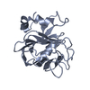
| Title | Bovine naive ultralong antibody AbD08 collected at 293K |
|---|
 Components Components | |
|---|
 Keywords Keywords | IMMUNE SYSTEM / ultralong / immunoglobulin / naive |
|---|
| Biological species |   Bos taurus (domestic cattle) Bos taurus (domestic cattle) |
|---|
| Method |  X-RAY DIFFRACTION / SYNCHROTRON / MOLECULAR REPLACEMENT / Resolution: 2.2 Å X-RAY DIFFRACTION / SYNCHROTRON / MOLECULAR REPLACEMENT / Resolution: 2.2 Å |
|---|
 Authors Authors | Clarke, J.D. / Mikolajek, H. / Stuart, D.I. / Owens, R.J. |
|---|
| Funding support |  United Kingdom, 1items United Kingdom, 1items | Organization | Grant number | Country |
|---|
| Biotechnology and Biological Sciences Research Council (BBSRC) | BB/M011224/1 | United Kingdom |
|
|---|
 Citation Citation | Journal: Iucrj / Year: 2023Title: Protein-to-structure pipeline for ambient-temperature in situ crystallography at VMXi. Authors: Mikolajek, H. / Sanchez-Weatherby, J. / Sandy, J. / Gildea, R.J. / Campeotto, I. / Cheruvara, H. / Clarke, J.D. / Foster, T. / Fujii, S. / Paulsen, I.T. / Shah, B.S. / Hough, M.A. |
|---|
| History | | Deposition | Feb 9, 2023 | Deposition site: PDBE / Processing site: PDBE |
|---|
| Revision 1.0 | May 24, 2023 | Provider: repository / Type: Initial release |
|---|
| Revision 1.1 | May 31, 2023 | Group: Database references / Category: citation / citation_author
Item: _citation.pdbx_database_id_PubMed / _citation.title / _citation_author.identifier_ORCID |
|---|
| Revision 1.2 | Jul 19, 2023 | Group: Data collection / Database references / Category: citation / diffrn_source
Item: _citation.journal_volume / _citation.page_first ..._citation.journal_volume / _citation.page_first / _citation.page_last / _diffrn_source.pdbx_synchrotron_site |
|---|
| Revision 1.3 | Oct 16, 2024 | Group: Data collection / Structure summary
Category: chem_comp_atom / chem_comp_bond ...chem_comp_atom / chem_comp_bond / diffrn_source / pdbx_entry_details / pdbx_modification_feature
Item: _diffrn_source.pdbx_synchrotron_site |
|---|
|
|---|
 Movie
Movie Controller


 Open data
 Basic information
 Structure visualization
 Molmil
 Downloads & links
 Other downloads


 PDBj
PDBj

 Assembly
 Homo sapiens (human)
 Mass: 18.015 Da / Num. of mol.: 52 / Source method: isolated from a natural source / Formula: H2O
 Sample preparation