 Movie
Movie Controller


+ Open data

- Basic information
| Entry | Database: PDB / ID: 8ch1 | ||||||
|---|---|---|---|---|---|---|---|
| Title | PBP AccA from A. vitis S4 in complex with Agrocinopine A | ||||||
 Components Components | Agrocinopine utilization periplasmic binding protein AccA | ||||||
 Keywords Keywords | TRANSPORT PROTEIN / periplasmic binding protein / solute binding protein | ||||||
| Function / homology | : / : / 2-O-phosphono-alpha-L-arabinopyranose / DI(HYDROXYETHYL)ETHER / 2-O-phosphono-beta-L-arabinopyranose Function and homology information Function and homology information | ||||||
| Biological species |  Agrobacterium vitis S4 (bacteria) Agrobacterium vitis S4 (bacteria) | ||||||
| Method |  X-RAY DIFFRACTION / SYNCHROTRON / MOLECULAR REPLACEMENT / Resolution: 1.496 Å X-RAY DIFFRACTION / SYNCHROTRON / MOLECULAR REPLACEMENT / Resolution: 1.496 Å | ||||||
 Authors Authors | Morera, S. / Vigouroux, A. | ||||||
| Funding support | 1items
| ||||||
 Citation Citation | Journal: Biochem.J. / Year: 2024 Title: A highly conserved ligand-binding site for AccA transporters of antibiotic and quorum-sensing regulator in Agrobacterium leads to a different specificity. Authors: Morera, S. / Vigouroux, A. / Aumont-Nicaise, M. / Ahmar, M. / Meyer, T. / El Sahili, A. / Deicsics, G. / Gonzalez-Mula, A. / Li, S. / Dore, J. / Sirigu, S. / Legrand, P. / Penot, C. / Andre, ...Authors: Morera, S. / Vigouroux, A. / Aumont-Nicaise, M. / Ahmar, M. / Meyer, T. / El Sahili, A. / Deicsics, G. / Gonzalez-Mula, A. / Li, S. / Dore, J. / Sirigu, S. / Legrand, P. / Penot, C. / Andre, F. / Faure, D. / Soulere, L. / Queneau, Y. / Vial, L. | ||||||
| History |
|
- Structure visualization
| Structure viewer | Molecule: MolmilJmol/JSmol |
|---|
- Downloads & links
-Download
| PDBx/mmCIF format | 8ch1.cif.gz | 297.3 KB | Display | PDBx/mmCIF format |
|---|---|---|---|---|
| PDB format | pdb8ch1.ent.gz | 242.5 KB | Display | PDB format |
| PDBx/mmJSON format | 8ch1.json.gz | Tree view | PDBx/mmJSON format | |
| Others |  Other downloads Other downloads |
-Validation report
| Arichive directory | https://data.pdbj.org/pub/pdb/validation_reports/ch/8ch1ftp://data.pdbj.org/pub/pdb/validation_reports/ch/8ch1 | HTTPS FTP |
|---|
-Related structure data
| Related structure data |  8c6rC  8c6uC  8c6wC 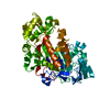 8c6yC  8c75C  8cawC  8cayC 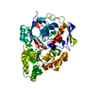 8cb9C  8cdoC 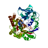 8ch2C 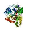 8ch3C 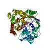 8chcC  8ci6C 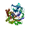 8cjuC 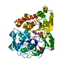 8ckdC  8ckeC 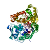 8ckoC C: citing same article ( |
|---|---|
| Similar structure data |
-Links
 PDBj
PDBj- Assembly
| Deposited unit | 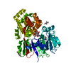
| ||||||||
|---|---|---|---|---|---|---|---|---|---|
| 1 |
| ||||||||
| Unit cell |
|
-Components
-Protein , 1 types, 1 molecules A
| #1: Protein | Mass: 56652.438 Da / Num. of mol.: 1 Source method: isolated from a genetically manipulated source Source: (gene. exp.) Agrobacterium vitis S4 (bacteria)Production host: |
|---|
-Sugars , 3 types, 4 molecules 
| #2: Polysaccharide | Source method: isolated from a genetically manipulated source Details: phosphodiester of sucrose and L-arabinose / References: BIRD: PRD_002473 #3: Sugar | ChemComp-VDF / |  Type: L-saccharide, beta linking, Oligosaccharide / Class: Nutrient / Mass: 230.110 Da / Num. of mol.: 1 / Source method: obtained synthetically / Formula: C5H11O8P / Details: phosphodiester of sucrose and L-arabinose / Feature type: SUBJECT OF INVESTIGATION / References: BIRD: PRD_002473 Type: L-saccharide, beta linking, Oligosaccharide / Class: Nutrient / Mass: 230.110 Da / Num. of mol.: 1 / Source method: obtained synthetically / Formula: C5H11O8P / Details: phosphodiester of sucrose and L-arabinose / Feature type: SUBJECT OF INVESTIGATION / References: BIRD: PRD_002473#4: Sugar | ChemComp-LAO / |  Type: L-saccharide, alpha linking, Oligosaccharide / Class: Nutrient / Mass: 230.110 Da / Num. of mol.: 1 / Source method: obtained synthetically / Formula: C5H11O8P / Details: phosphodiester of sucrose and L-arabinose / Feature type: SUBJECT OF INVESTIGATION / References: BIRD: PRD_002470 Type: L-saccharide, alpha linking, Oligosaccharide / Class: Nutrient / Mass: 230.110 Da / Num. of mol.: 1 / Source method: obtained synthetically / Formula: C5H11O8P / Details: phosphodiester of sucrose and L-arabinose / Feature type: SUBJECT OF INVESTIGATION / References: BIRD: PRD_002470 |
|---|
-Non-polymers , 3 types, 455 molecules 


| #5: Chemical |  Mass: 62.068 Da / Num. of mol.: 3 / Source method: obtained synthetically / Formula: C2H6O2 Mass: 62.068 Da / Num. of mol.: 3 / Source method: obtained synthetically / Formula: C2H6O2#6: Chemical | ChemComp-PEG / |  Mass: 106.120 Da / Num. of mol.: 1 / Source method: obtained synthetically / Formula: C4H10O3 Mass: 106.120 Da / Num. of mol.: 1 / Source method: obtained synthetically / Formula: C4H10O3#7: Water | ChemComp-HOH / | Mass: 18.015 Da / Num. of mol.: 451 / Source method: isolated from a natural source / Formula: H2O |
|---|
-Details
| Compound details | A member of the class of agrocinopines that consists of sucrose and L-arabinose units joined via a ...A member of the class of agrocinopines that consists of sucrose and L-arabinose units joined via a phosphodiester linkage between position 4F of sucrose and position 2 of arabinose. |
|---|---|
| Has ligand of interest | Y |
-Experimental details
-Experiment
| Experiment | Method: X-RAY DIFFRACTION / Number of used crystals: 1 |
|---|
- Sample preparation
| Crystal | Density Matthews: 1.91 Å3/Da / Density % sol: 35.46 % |
|---|---|
| Crystal grow | Temperature: 292 K / Method: vapor diffusion, hanging drop / pH: 7 / Details: PEG 8000, HEPES |
-Data collection
| Diffraction | Mean temperature: 100 K / Serial crystal experiment: N |
|---|---|
| Diffraction source | Source: SYNCHROTRON / Site: SOLEIL  / Beamline: PROXIMA 2 / Wavelength: 0.97933 Å / Beamline: PROXIMA 2 / Wavelength: 0.97933 Å |
| Detector | Type: DECTRIS EIGER X 9M / Detector: PIXEL / Date: Jun 29, 2016 |
| Radiation | Protocol: SINGLE WAVELENGTH / Monochromatic (M) / Laue (L): M / Scattering type: x-ray |
| Radiation wavelength | Wavelength: 0.97933 Å / Relative weight: 1 |
| Reflection | Resolution: 1.496→47.3 Å / Num. obs: 70531 / % possible obs: 99.5 % / Redundancy: 6.6 % / CC1/2: 0.99 / Rmerge(I) obs: 0.09 / Net I/σ(I): 15.9 |
| Reflection shell | Resolution: 1.496→1.59 Å / Rmerge(I) obs: 1.303 / Num. unique obs: 10968 / CC1/2: 0.97 |
- Processing
| Software |
| ||||||||||||||||||||||||||||||||||||||||||||||||||||||||||||
|---|---|---|---|---|---|---|---|---|---|---|---|---|---|---|---|---|---|---|---|---|---|---|---|---|---|---|---|---|---|---|---|---|---|---|---|---|---|---|---|---|---|---|---|---|---|---|---|---|---|---|---|---|---|---|---|---|---|---|---|---|---|
| Refinement | Method to determine structure: MOLECULAR REPLACEMENT / Resolution: 1.496→47.3 Å / Cor.coef. Fo:Fc: 0.962 / Cor.coef. Fo:Fc free: 0.955 / SU R Cruickshank DPI: 0.088 / Cross valid method: THROUGHOUT / SU R Blow DPI: 0.079 / SU Rfree Blow DPI: 0.078 / SU Rfree Cruickshank DPI: 0.075
| ||||||||||||||||||||||||||||||||||||||||||||||||||||||||||||
| Displacement parameters | Biso mean: 27.8 Å2
| ||||||||||||||||||||||||||||||||||||||||||||||||||||||||||||
| Refine analyze | Luzzati coordinate error obs: 0.18 Å | ||||||||||||||||||||||||||||||||||||||||||||||||||||||||||||
| Refinement step | Cycle: LAST / Resolution: 1.496→47.3 Å
| ||||||||||||||||||||||||||||||||||||||||||||||||||||||||||||
| Refine LS restraints |
| ||||||||||||||||||||||||||||||||||||||||||||||||||||||||||||
| LS refinement shell | Resolution: 1.496→1.51 Å
| ||||||||||||||||||||||||||||||||||||||||||||||||||||||||||||
| Refinement TLS params. | Origin x: 17.6356 Å / Origin y: -1.327 Å / Origin z: -19.1469 Å
| ||||||||||||||||||||||||||||||||||||||||||||||||||||||||||||
| Refinement TLS group | Selection details: { A|* } |
