 Movie
Movie Controller

[English] 日本語
 Yorodumi
- PDB-8cf7: Dimethylated RSL-R5 in complex with cucurbit[7]uril, C121 sheet a... -
+ Open data

- Basic information
| Entry | Database: PDB / ID: 8cf7 | |||||||||
|---|---|---|---|---|---|---|---|---|---|---|
| Title | Dimethylated RSL-R5 in complex with cucurbit[7]uril, C121 sheet assembly | |||||||||
 Components Components | RSL-R5 | |||||||||
 Keywords Keywords | SUGAR BINDING PROTEIN / lectin / cucurbituril / biomaterials / supramolecular | |||||||||
| Function / homology | cucurbit[7]uril Function and homology information Function and homology information | |||||||||
| Biological species |  Ralstonia solanacearum (bacteria) Ralstonia solanacearum (bacteria) | |||||||||
| Method |  X-RAY DIFFRACTION / SYNCHROTRON / MOLECULAR REPLACEMENT / Resolution: 1.14 Å X-RAY DIFFRACTION / SYNCHROTRON / MOLECULAR REPLACEMENT / Resolution: 1.14 Å | |||||||||
 Authors Authors | Ramberg, K. / Crowley, P.B. | |||||||||
| Funding support |  Ireland, 2items Ireland, 2items
| |||||||||
 Citation Citation | Journal: J.Struct.Biol. / Year: 2023 Title: Cage versus sheet: Probing the Determinants of Protein - Cucurbit[7]uril Crystalline Architectures. Authors: Ramberg, K.O. / Crowley, P.B. | |||||||||
| History |
|
- Structure visualization
| Structure viewer | Molecule: MolmilJmol/JSmol |
|---|
- Downloads & links
-Download
| PDBx/mmCIF format | 8cf7.cif.gz | 152.4 KB | Display | PDBx/mmCIF format |
|---|---|---|---|---|
| PDB format | pdb8cf7.ent.gz | 127.5 KB | Display | PDB format |
| PDBx/mmJSON format | 8cf7.json.gz | Tree view | PDBx/mmJSON format | |
| Others |  Other downloads Other downloads |
-Validation report
| Arichive directory | https://data.pdbj.org/pub/pdb/validation_reports/cf/8cf7ftp://data.pdbj.org/pub/pdb/validation_reports/cf/8cf7 | HTTPS FTP |
|---|
-Related structure data

-Links
 PDBj
PDBj
- Assembly
| Deposited unit | 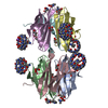
| ||||||||
|---|---|---|---|---|---|---|---|---|---|
| 1 | 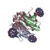
| ||||||||
| 2 | 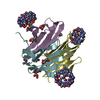
| ||||||||
| Unit cell |
| ||||||||
| Components on special symmetry positions |
|
-Components
| #1: Protein | Mass: 9844.689 Da / Num. of mol.: 6 Source method: isolated from a genetically manipulated source Source: (gene. exp.) Ralstonia solanacearum (bacteria) / Production host: #2: Chemical | ChemComp-QQ7 /   Mass: 1162.962 Da / Num. of mol.: 6 / Source method: obtained synthetically / Formula: C42H42N28O14 / Feature type: SUBJECT OF INVESTIGATION Mass: 1162.962 Da / Num. of mol.: 6 / Source method: obtained synthetically / Formula: C42H42N28O14 / Feature type: SUBJECT OF INVESTIGATION#3: Chemical | ChemComp-GOL /   Mass: 92.094 Da / Num. of mol.: 15 / Source method: obtained synthetically / Formula: C3H8O3 Mass: 92.094 Da / Num. of mol.: 15 / Source method: obtained synthetically / Formula: C3H8O3#4: Chemical |   Mass: 22.990 Da / Num. of mol.: 2 / Source method: obtained synthetically / Formula: Na Mass: 22.990 Da / Num. of mol.: 2 / Source method: obtained synthetically / Formula: Na#5: Water | ChemComp-HOH / |  Mass: 18.015 Da / Num. of mol.: 929 / Source method: isolated from a natural source / Formula: H2O Mass: 18.015 Da / Num. of mol.: 929 / Source method: isolated from a natural source / Formula: H2OHas ligand of interest | Y | |
|---|
-Experimental details
-Experiment
| Experiment | Method: X-RAY DIFFRACTION / Number of used crystals: 1 |
|---|
- Sample preparation
| Crystal | Density Matthews: 2.62 Å3/Da / Density % sol: 54 % / Description: hexagonal plate |
|---|---|
| Crystal grow | Temperature: 293.15 K / Method: vapor diffusion / Details: 25 % PEG3350 0.1 M Bis-Tris pH 5.5 0.2 M NaCl |
-Data collection
| Diffraction | Mean temperature: 100 K / Serial crystal experiment: N |
|---|---|
| Diffraction source | Source: SYNCHROTRON / Site: SOLEIL  / Beamline: PROXIMA 2 / Wavelength: 0.98 Å / Beamline: PROXIMA 2 / Wavelength: 0.98 Å |
| Detector | Type: DECTRIS EIGER X 9M / Detector: PIXEL / Date: Jul 25, 2020 |
| Radiation | Protocol: SINGLE WAVELENGTH / Monochromatic (M) / Laue (L): M / Scattering type: x-ray |
| Radiation wavelength | Wavelength: 0.98 Å / Relative weight: 1 |
| Reflection | Resolution: 1.14→46.74 Å / Num. obs: 212450 / % possible obs: 95.1 % / Redundancy: 4.9 % / Biso Wilson estimate: 12.02 Å2 / CC1/2: 0.99 / Rpim(I) all: 0.017 / Rrim(I) all: 0.044 / Net I/σ(I): 18.2 |
| Reflection shell | Resolution: 1.14→1.16 Å / Redundancy: 33.7 % / Mean I/σ(I) obs: 2.1 / Num. unique obs: 7323 / CC1/2: 0.93 / Rpim(I) all: 0.256 / Rrim(I) all: 0.465 / % possible all: 66.3 |
- Processing
| Software |
| |||||||||||||||||||||||||||||||||||||||||||||||||||||||||||||||||||||||||||||||||||||||||||||||||||||||||||||||||||||||||||||||||||||||||||||||||||||||||||||||||||||||||||||||||||||||||||||||||||||||||||||||||||||||||
|---|---|---|---|---|---|---|---|---|---|---|---|---|---|---|---|---|---|---|---|---|---|---|---|---|---|---|---|---|---|---|---|---|---|---|---|---|---|---|---|---|---|---|---|---|---|---|---|---|---|---|---|---|---|---|---|---|---|---|---|---|---|---|---|---|---|---|---|---|---|---|---|---|---|---|---|---|---|---|---|---|---|---|---|---|---|---|---|---|---|---|---|---|---|---|---|---|---|---|---|---|---|---|---|---|---|---|---|---|---|---|---|---|---|---|---|---|---|---|---|---|---|---|---|---|---|---|---|---|---|---|---|---|---|---|---|---|---|---|---|---|---|---|---|---|---|---|---|---|---|---|---|---|---|---|---|---|---|---|---|---|---|---|---|---|---|---|---|---|---|---|---|---|---|---|---|---|---|---|---|---|---|---|---|---|---|---|---|---|---|---|---|---|---|---|---|---|---|---|---|---|---|---|---|---|---|---|---|---|---|---|---|---|---|---|---|---|---|---|
| Refinement | Method to determine structure: MOLECULAR REPLACEMENT / Resolution: 1.14→46.74 Å / SU ML: 0.1 / Cross valid method: FREE R-VALUE / σ(F): 1.35 / Phase error: 19.44 / Stereochemistry target values: ML
| |||||||||||||||||||||||||||||||||||||||||||||||||||||||||||||||||||||||||||||||||||||||||||||||||||||||||||||||||||||||||||||||||||||||||||||||||||||||||||||||||||||||||||||||||||||||||||||||||||||||||||||||||||||||||
| Solvent computation | Shrinkage radii: 0.9 Å / VDW probe radii: 1.11 Å / Solvent model: FLAT BULK SOLVENT MODEL | |||||||||||||||||||||||||||||||||||||||||||||||||||||||||||||||||||||||||||||||||||||||||||||||||||||||||||||||||||||||||||||||||||||||||||||||||||||||||||||||||||||||||||||||||||||||||||||||||||||||||||||||||||||||||
| Refinement step | Cycle: LAST / Resolution: 1.14→46.74 Å
| |||||||||||||||||||||||||||||||||||||||||||||||||||||||||||||||||||||||||||||||||||||||||||||||||||||||||||||||||||||||||||||||||||||||||||||||||||||||||||||||||||||||||||||||||||||||||||||||||||||||||||||||||||||||||
| Refine LS restraints |
| |||||||||||||||||||||||||||||||||||||||||||||||||||||||||||||||||||||||||||||||||||||||||||||||||||||||||||||||||||||||||||||||||||||||||||||||||||||||||||||||||||||||||||||||||||||||||||||||||||||||||||||||||||||||||
| LS refinement shell |
|
