 Movie
Movie Controller

[English] 日本語
 Yorodumi
- PDB-8cdf: Structure of glutarate hydroxylase (GlaH) from Escherichia coli a... -
+ Open data

- Basic information
| Entry | Database: PDB / ID: 8cdf | ||||||
|---|---|---|---|---|---|---|---|
| Title | Structure of glutarate hydroxylase (GlaH) from Escherichia coli at a resolution of 1.8 angstrom obtained as a contaminant during routine use of E. coli as an expression host | ||||||
 Components Components | Glutarate 2-hydroxylase | ||||||
 Keywords Keywords | METAL BINDING PROTEIN / Carbon-starvation induced / hydroxylase / non haem Fe (II)-dependent oxygenase | ||||||
| Function / homology |  Function and homology information Function and homology informationglutarate dioxygenase / oxidoreductase activity, acting on paired donors, with incorporation or reduction of molecular oxygen, with 2-oxoglutarate as one donor, and the other dehydrogenated / glutarate dioxygenase activity / L-lysine catabolic process / ferrous iron binding Similarity search - Function | ||||||
| Biological species |  | ||||||
| Method |  X-RAY DIFFRACTION / SYNCHROTRON / MOLECULAR REPLACEMENT / Resolution: 1.77 Å X-RAY DIFFRACTION / SYNCHROTRON / MOLECULAR REPLACEMENT / Resolution: 1.77 Å | ||||||
 Authors Authors | Adeyeye, A.A. / Schubert, W. | ||||||
| Funding support |  South Africa, 1items South Africa, 1items
| ||||||
 Citation Citation | Journal: To Be Published Title: Structure of glutarate hydroxylase (GlaH) from Escherichia coli at a resolution of 1.8 angstrom obtained as a contaminant during routine use of E. coli as an expression host Authors: Adeyeye, A.A. / Schubert, W. | ||||||
| History |
|
- Structure visualization
| Structure viewer | Molecule: MolmilJmol/JSmol |
|---|
- Downloads & links
-Download
| PDBx/mmCIF format | 8cdf.cif.gz | 95.8 KB | Display | PDBx/mmCIF format |
|---|---|---|---|---|
| PDB format | pdb8cdf.ent.gz | 57.8 KB | Display | PDB format |
| PDBx/mmJSON format | 8cdf.json.gz | Tree view | PDBx/mmJSON format | |
| Others |  Other downloads Other downloads |
-Validation report
| Arichive directory | https://data.pdbj.org/pub/pdb/validation_reports/cd/8cdfftp://data.pdbj.org/pub/pdb/validation_reports/cd/8cdf | HTTPS FTP |
|---|
-Related structure data
| Similar structure data |
|---|
-Links
 PDBj
PDBj


- Assembly
| Deposited unit | 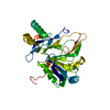
| ||||||||||||
|---|---|---|---|---|---|---|---|---|---|---|---|---|---|
| 1 | 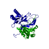
| ||||||||||||
| Unit cell |
| ||||||||||||
| Components on special symmetry positions |
|
-Components
| #1: Protein | Mass: 35974.762 Da / Num. of mol.: 1 Source method: isolated from a genetically manipulated source Source: (gene. exp.) Gene: csiD, glaH, ACU57_06710, AM464_04980, BGM66_004646, BJI68_04905, C5N07_05545, CTR35_002510, EIZ93_05365, EL79_1036, FOI11_00325, FOI11_022855, FV293_23215, FWK02_21895, G3V95_09995, GIB53_ ...Gene: csiD, glaH, ACU57_06710, AM464_04980, BGM66_004646, BJI68_04905, C5N07_05545, CTR35_002510, EIZ93_05365, EL79_1036, FOI11_00325, FOI11_022855, FV293_23215, FWK02_21895, G3V95_09995, GIB53_09245, GJO56_18475, GKF89_16305, GNW61_23135, GRW05_29650, GRW24_16590, GRW56_10550, GRW57_12335, HMV95_03335, J0541_002578, JNP96_20920, NCTC9045_01367, NCTC9073_04720, NCTC9117_01751, SAMEA3751407_03408 Production host: |
|---|---|
| #2: Chemical | ChemComp-FE2 /   Mass: 55.845 Da / Num. of mol.: 1 / Source method: obtained synthetically / Formula: Fe / Feature type: SUBJECT OF INVESTIGATION Mass: 55.845 Da / Num. of mol.: 1 / Source method: obtained synthetically / Formula: Fe / Feature type: SUBJECT OF INVESTIGATION |
| #3: Water | ChemComp-HOH /  Mass: 18.015 Da / Num. of mol.: 193 / Source method: isolated from a natural source / Formula: H2O Mass: 18.015 Da / Num. of mol.: 193 / Source method: isolated from a natural source / Formula: H2O |
| Has ligand of interest | Y |
-Experimental details
-Experiment
| Experiment | Method: X-RAY DIFFRACTION / Number of used crystals: 1 |
|---|
- Sample preparation
| Crystal | Density Matthews: 3.48 Å3/Da / Density % sol: 64.64 % |
|---|---|
| Crystal grow | Temperature: 291.15 K / Method: vapor diffusion, hanging drop / pH: 6.9 / Details: 47 mM K3PO4, 0.4 M (NH4)2SO4, 200 mM KCl, 1mM EDTA |
-Data collection
| Diffraction | Mean temperature: 100 K / Serial crystal experiment: N |
|---|---|
| Diffraction source | Source: SYNCHROTRON / Site: Diamond  / Beamline: I04 / Wavelength: 0.9795 Å / Beamline: I04 / Wavelength: 0.9795 Å |
| Detector | Type: DECTRIS EIGER X 16M / Detector: PIXEL / Date: Jul 3, 2020 |
| Radiation | Protocol: SINGLE WAVELENGTH / Monochromatic (M) / Laue (L): M / Scattering type: x-ray |
| Radiation wavelength | Wavelength: 0.9795 Å / Relative weight: 1 |
| Reflection | Resolution: 1.77→53.37 Å / Num. obs: 49508 / % possible obs: 99.95 % / Redundancy: 11.9 % / Biso Wilson estimate: 33.99 Å2 / CC1/2: 0.999 / Rmerge(I) obs: 0.102 / Rrim(I) all: 0.106 / Net I/σ(I): 11.9 |
| Reflection shell | Resolution: 1.77→1.8 Å / Redundancy: 6 % / Mean I/σ(I) obs: 0.3 / Num. unique obs: 2412 / CC1/2: 0.4 / % possible all: 99.1 |
- Processing
| Software |
| |||||||||||||||||||||||||||||||||||||||||||||||||||||||||||||||||||||||||||||||||||||||||||||||||||||||||
|---|---|---|---|---|---|---|---|---|---|---|---|---|---|---|---|---|---|---|---|---|---|---|---|---|---|---|---|---|---|---|---|---|---|---|---|---|---|---|---|---|---|---|---|---|---|---|---|---|---|---|---|---|---|---|---|---|---|---|---|---|---|---|---|---|---|---|---|---|---|---|---|---|---|---|---|---|---|---|---|---|---|---|---|---|---|---|---|---|---|---|---|---|---|---|---|---|---|---|---|---|---|---|---|---|---|---|
| Refinement | Method to determine structure: MOLECULAR REPLACEMENT / Resolution: 1.77→50.35 Å / SU ML: 0.316 / Cross valid method: FREE R-VALUE / σ(F): 1.33 / Phase error: 26.5487 Stereochemistry target values: GeoStd + Monomer Library + CDL v1.2
| |||||||||||||||||||||||||||||||||||||||||||||||||||||||||||||||||||||||||||||||||||||||||||||||||||||||||
| Solvent computation | Shrinkage radii: 0.9 Å / VDW probe radii: 1.11 Å / Solvent model: FLAT BULK SOLVENT MODEL | |||||||||||||||||||||||||||||||||||||||||||||||||||||||||||||||||||||||||||||||||||||||||||||||||||||||||
| Displacement parameters | Biso mean: 39.05 Å2 | |||||||||||||||||||||||||||||||||||||||||||||||||||||||||||||||||||||||||||||||||||||||||||||||||||||||||
| Refinement step | Cycle: LAST / Resolution: 1.77→50.35 Å
| |||||||||||||||||||||||||||||||||||||||||||||||||||||||||||||||||||||||||||||||||||||||||||||||||||||||||
| Refine LS restraints |
| |||||||||||||||||||||||||||||||||||||||||||||||||||||||||||||||||||||||||||||||||||||||||||||||||||||||||
| LS refinement shell |
|
