 Movie
Movie Controller

[English] 日本語
 Yorodumi
- PDB-8cb5: The Transcriptional Regulator PrfA from Listeria Monocytogenes in... -
+ Open data

- Basic information
| Entry | Database: PDB / ID: 8cb5 | |||||||||
|---|---|---|---|---|---|---|---|---|---|---|
| Title | The Transcriptional Regulator PrfA from Listeria Monocytogenes in complex with tripeptide Glu-Val-Phe | |||||||||
 Components Components |
| |||||||||
 Keywords Keywords | TRANSCRIPTION / DNA BINDING PROTEIN / PRFA / LISTERIA INHIBITION / VIRULENCE | |||||||||
| Function / homology |  Function and homology information Function and homology information | |||||||||
| Biological species |  Listeria monocytogenes (bacteria) Listeria monocytogenes (bacteria) | |||||||||
| Method |  X-RAY DIFFRACTION / SYNCHROTRON / MOLECULAR REPLACEMENT / Resolution: 2.25 Å X-RAY DIFFRACTION / SYNCHROTRON / MOLECULAR REPLACEMENT / Resolution: 2.25 Å | |||||||||
 Authors Authors | Oelker, M. / Sauer-Eriksson, A.E. | |||||||||
| Funding support |  Sweden, 2items Sweden, 2items
| |||||||||
 Citation Citation | Journal: Cell Rep / Year: 2025 Title: Structural basis of promiscuous inhibition of Listeria virulence activator PrfA by oligopeptides. Authors: Hainzl, T. / Scortti, M. / Lindgren, C. / Grundstrom, C. / Krypotou, E. / Vazquez-Boland, J.A. / Sauer-Eriksson, A.E. #1: Journal: Cell Rep / Year: 2019Title: Control of Bacterial Virulence through the Peptide Signature of the Habitat. Authors: Krypotou, E. / Scortti, M. / Grundstrom, C. / Oelker, M. / Luisi, B.F. / Sauer-Eriksson, A.E. / Vazquez-Boland, J. | |||||||||
| History |
|
- Structure visualization
| Structure viewer | Molecule: MolmilJmol/JSmol |
|---|
- Downloads & links
-Download
| PDBx/mmCIF format | 8cb5.cif.gz | 107.7 KB | Display | PDBx/mmCIF format |
|---|---|---|---|---|
| PDB format | pdb8cb5.ent.gz | 81.1 KB | Display | PDB format |
| PDBx/mmJSON format | 8cb5.json.gz | Tree view | PDBx/mmJSON format | |
| Others |  Other downloads Other downloads |
-Validation report
| Arichive directory | https://data.pdbj.org/pub/pdb/validation_reports/cb/8cb5ftp://data.pdbj.org/pub/pdb/validation_reports/cb/8cb5 | HTTPS FTP |
|---|
-Related structure data
| Related structure data |  8cb4C  8cb7C  8cb8C  8cbgC 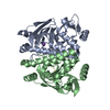 8cbiC 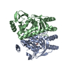 8cbpC C: citing same article ( |
|---|---|
| Similar structure data |
-Links
 PDBj
PDBj


- Assembly
| Deposited unit | 
| ||||||||
|---|---|---|---|---|---|---|---|---|---|
| 1 |
| ||||||||
| Unit cell |
|
-Components
| #1: Protein | Mass: 27457.521 Da / Num. of mol.: 2 Source method: isolated from a genetically manipulated source Source: (gene. exp.) Listeria monocytogenes (bacteria) / Gene: prfA, lmo0200 / Production host: #2: Protein/peptide | Mass: 393.434 Da / Num. of mol.: 2 / Source method: obtained synthetically / Source: (synth.) Listeria monocytogenes (bacteria)#3: Chemical |   Mass: 60.095 Da / Num. of mol.: 3 / Source method: obtained synthetically / Formula: C3H8O Mass: 60.095 Da / Num. of mol.: 3 / Source method: obtained synthetically / Formula: C3H8O#4: Chemical |   Mass: 22.990 Da / Num. of mol.: 2 / Source method: obtained synthetically / Formula: Na Mass: 22.990 Da / Num. of mol.: 2 / Source method: obtained synthetically / Formula: Na#5: Water | ChemComp-HOH / |  Mass: 18.015 Da / Num. of mol.: 53 / Source method: isolated from a natural source / Formula: H2O Mass: 18.015 Da / Num. of mol.: 53 / Source method: isolated from a natural source / Formula: H2OHas ligand of interest | N | Has protein modification | N | |
|---|
-Experimental details
-Experiment
| Experiment | Method: X-RAY DIFFRACTION / Number of used crystals: 1 |
|---|
- Sample preparation
| Crystal | Density Matthews: 2.13 Å3/Da / Density % sol: 42.18 % |
|---|---|
| Crystal grow | Temperature: 291 K / Method: vapor diffusion, sitting drop / pH: 5.5 Details: Protein: 3.1-6.6 mg mL-1, 20 mM NaP pH 6.5, 200 mM NaCl was mixed with peptides (0.5-2.5% DMSO, 1-4 mM DTT) in ratio 1:10. Well solution: 100 mM Nacitrate pH 5.1-5.7, 17% isopropanol, 16-26% ...Details: Protein: 3.1-6.6 mg mL-1, 20 mM NaP pH 6.5, 200 mM NaCl was mixed with peptides (0.5-2.5% DMSO, 1-4 mM DTT) in ratio 1:10. Well solution: 100 mM Nacitrate pH 5.1-5.7, 17% isopropanol, 16-26% PEG4000. Dropsize 1:1 microL Cryoprotection: 35% PEG4000 PH range: 5.1-5.7 |
-Data collection
| Diffraction | Mean temperature: 100 K / Serial crystal experiment: N |
|---|---|
| Diffraction source | Source: SYNCHROTRON / Site: MAX IV / Beamline: BioMAX / Wavelength: 0.9537 Å |
| Detector | Type: DECTRIS EIGER X 16M / Detector: PIXEL / Date: Mar 22, 2019 |
| Radiation | Protocol: SINGLE WAVELENGTH / Monochromatic (M) / Laue (L): M / Scattering type: x-ray |
| Radiation wavelength | Wavelength: 0.9537 Å / Relative weight: 1 |
| Reflection | Resolution: 2.25→47.33 Å / Num. obs: 23242 / % possible obs: 100 % / Redundancy: 12.7 % / Biso Wilson estimate: 53.6 Å2 / CC1/2: 0.999 / Rpim(I) all: 0.053 / Net I/σ(I): 12.9 |
| Reflection shell | Resolution: 2.25→2.33 Å / Redundancy: 13.4 % / Mean I/σ(I) obs: 1.3 / Num. unique obs: 2263 / CC1/2: 0.546 / Rpim(I) all: 0.59 / % possible all: 100 |
- Processing
| Software |
| ||||||||||||||||||||||||||||||||||||||||||||||||||||||
|---|---|---|---|---|---|---|---|---|---|---|---|---|---|---|---|---|---|---|---|---|---|---|---|---|---|---|---|---|---|---|---|---|---|---|---|---|---|---|---|---|---|---|---|---|---|---|---|---|---|---|---|---|---|---|---|
| Refinement | Method to determine structure: MOLECULAR REPLACEMENT / Resolution: 2.25→47.33 Å / SU ML: 0.31 / Cross valid method: THROUGHOUT / σ(F): 1.35 / Phase error: 28.91 / Stereochemistry target values: ML
| ||||||||||||||||||||||||||||||||||||||||||||||||||||||
| Solvent computation | Shrinkage radii: 0.9 Å / VDW probe radii: 1.11 Å / Solvent model: FLAT BULK SOLVENT MODEL | ||||||||||||||||||||||||||||||||||||||||||||||||||||||
| Displacement parameters | Biso max: 140.64 Å2 / Biso mean: 69.4232 Å2 / Biso min: 33.58 Å2 | ||||||||||||||||||||||||||||||||||||||||||||||||||||||
| Refinement step | Cycle: final / Resolution: 2.25→47.33 Å
| ||||||||||||||||||||||||||||||||||||||||||||||||||||||
| LS refinement shell | Refine-ID: X-RAY DIFFRACTION / Rfactor Rfree error: 0 / Total num. of bins used: 8 / % reflection obs: 100 %
|
