 Movie
Movie Controller

[English] 日本語
 Yorodumi
- PDB-8c8n: In situ structure of the Nitrosopumilus maritimus S-layer - Two-f... -
+ Open data

- Basic information
| Entry | Database: PDB / ID: 8c8n | |||||||||
|---|---|---|---|---|---|---|---|---|---|---|
| Title | In situ structure of the Nitrosopumilus maritimus S-layer - Two-fold symmetry (C2) | |||||||||
 Components Components | Cell surface protein | |||||||||
 Keywords Keywords | STRUCTURAL PROTEIN / Nmar_1547 S-layer | |||||||||
| Function / homology | Uncharacterized protein Function and homology information Function and homology information | |||||||||
| Biological species |  Nitrosopumilus maritimus SCM1 (archaea) Nitrosopumilus maritimus SCM1 (archaea) | |||||||||
| Method | ELECTRON MICROSCOPY / subtomogram averaging / cryo EM / Resolution: 3.4 Å | |||||||||
 Authors Authors | von Kuegelgen, A. / Bharat, T. | |||||||||
| Funding support |  United Kingdom, 2items United Kingdom, 2items
| |||||||||
 Citation Citation | Journal: Nature / Year: 2024 Title: Membraneless channels sieve cations in ammonia-oxidizing marine archaea. Authors: Andriko von Kügelgen / C Keith Cassidy / Sofie van Dorst / Lennart L Pagani / Christopher Batters / Zephyr Ford / Jan Löwe / Vikram Alva / Phillip J Stansfeld / Tanmay A M Bharat /   Abstract: Nitrosopumilus maritimus is an ammonia-oxidizing archaeon that is crucial to the global nitrogen cycle. A critical step for nitrogen oxidation is the entrapment of ammonium ions from a dilute marine ...Nitrosopumilus maritimus is an ammonia-oxidizing archaeon that is crucial to the global nitrogen cycle. A critical step for nitrogen oxidation is the entrapment of ammonium ions from a dilute marine environment at the cell surface and their subsequent channelling to the cell membrane of N. maritimus. Here we elucidate the structure of the molecular machinery responsible for this process, comprising the surface layer (S-layer), using electron cryotomography and subtomogram averaging from cells. We supplemented our in situ structure of the ammonium-binding S-layer array with a single-particle electron cryomicroscopy structure, revealing detailed features of this immunoglobulin-rich and glycan-decorated S-layer. Biochemical analyses showed strong ammonium binding by the cell surface, which was lost after S-layer disassembly. Sensitive bioinformatic analyses identified similar S-layers in many ammonia-oxidizing archaea, with conserved sequence and structural characteristics. Moreover, molecular simulations and structure determination of ammonium-enriched specimens enabled us to examine the cation-binding properties of the S-layer, revealing how it concentrates ammonium ions on its cell-facing side, effectively acting as a multichannel sieve on the cell membrane. This in situ structural study illuminates the biogeochemically essential process of ammonium binding and channelling, common to many marine microorganisms that are fundamental to the nitrogen cycle. | |||||||||
| History |
|
- Structure visualization
| Structure viewer | Molecule: MolmilJmol/JSmol |
|---|
- Downloads & links
-Download
| PDBx/mmCIF format | 8c8n.cif.gz | 535 KB | Display | PDBx/mmCIF format |
|---|---|---|---|---|
| PDB format | pdb8c8n.ent.gz | 361.7 KB | Display | PDB format |
| PDBx/mmJSON format | 8c8n.json.gz | Tree view | PDBx/mmJSON format | |
| Others |  Other downloads Other downloads |
-Validation report
| Arichive directory | https://data.pdbj.org/pub/pdb/validation_reports/c8/8c8nftp://data.pdbj.org/pub/pdb/validation_reports/c8/8c8n | HTTPS FTP |
|---|
-Related structure data
| Related structure data | 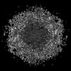 16487MC 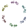 8c8kC  8c8lC 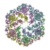 8c8mC  8c8oC 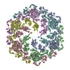 8c8rC M: map data used to model this data C: citing same article ( |
|---|---|
| Similar structure data |

-Links
 PDBj
PDBj- Assembly
| Deposited unit | 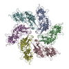
|
|---|---|
| 1 |
|
-Components
| #1: Protein | Mass: 183156.000 Da / Num. of mol.: 6 / Source method: isolated from a natural source / Source: (natural) Nitrosopumilus maritimus SCM1 (archaea) / Strain: SCM1 / References: UniProt: A9A4Y9Has protein modification | Y | |
|---|
-Experimental details
-Experiment
| Experiment | Method: ELECTRON MICROSCOPY |
|---|---|
| EM experiment | Aggregation state: CELL / 3D reconstruction method: subtomogram averaging |
- Sample preparation
| Component | Name: Nitrosopumilus maritimus S-layer / Type: ORGANELLE OR CELLULAR COMPONENT / Details: Nitrosopumilus maritimus S-layer C2 symmetrised / Entity ID: all / Source: NATURAL | ||||||||||||||||||||||||||||||||||||||||||||||||||||||||||||||||||||||||||||||||
|---|---|---|---|---|---|---|---|---|---|---|---|---|---|---|---|---|---|---|---|---|---|---|---|---|---|---|---|---|---|---|---|---|---|---|---|---|---|---|---|---|---|---|---|---|---|---|---|---|---|---|---|---|---|---|---|---|---|---|---|---|---|---|---|---|---|---|---|---|---|---|---|---|---|---|---|---|---|---|---|---|---|
| Molecular weight | Experimental value: NO | ||||||||||||||||||||||||||||||||||||||||||||||||||||||||||||||||||||||||||||||||
| Source (natural) | Organism: Nitrosopumilus maritimus SCM1 (archaea) / Strain: SCM1 / Cellular location: extracellular | ||||||||||||||||||||||||||||||||||||||||||||||||||||||||||||||||||||||||||||||||
| Buffer solution | pH: 7.4 / Details: Modified Syntheic Crenarchaeota medium | ||||||||||||||||||||||||||||||||||||||||||||||||||||||||||||||||||||||||||||||||
| Buffer component |
| ||||||||||||||||||||||||||||||||||||||||||||||||||||||||||||||||||||||||||||||||
| Specimen | Embedding applied: NO / Shadowing applied: NO / Staining applied: NO / Vitrification applied: YES / Details: Nitrosopumilus maritimus cells | ||||||||||||||||||||||||||||||||||||||||||||||||||||||||||||||||||||||||||||||||
| Specimen support | Details: 15 mA / Grid material: COPPER/RHODIUM / Grid mesh size: 200 divisions/in. / Grid type: Quantifoil R2/2 | ||||||||||||||||||||||||||||||||||||||||||||||||||||||||||||||||||||||||||||||||
| Vitrification | Instrument: FEI VITROBOT MARK IV / Cryogen name: NITROGEN / Humidity: 100 % / Chamber temperature: 283.15 K Details: absorption for 60 sec and blotted for 5 sec with blot force -10 |
- Electron microscopy imaging
| Experimental equipment |  Model: Titan Krios / Image courtesy: FEI Company |
|---|---|
| Microscopy | Model: FEI TITAN KRIOS |
| Electron gun | Electron source:  FIELD EMISSION GUN / Accelerating voltage: 300 kV / Illumination mode: FLOOD BEAM FIELD EMISSION GUN / Accelerating voltage: 300 kV / Illumination mode: FLOOD BEAM |
| Electron lens | Mode: BRIGHT FIELD / Nominal magnification: 105000 X / Calibrated magnification: 105000 X / Nominal defocus max: 5000 nm / Nominal defocus min: 2000 nm / Calibrated defocus min: 2000 nm / Calibrated defocus max: 5000 nm / Cs: 2.7 mm / C2 aperture diameter: 70 µm / Alignment procedure: COMA FREE |
| Specimen holder | Cryogen: NITROGEN / Specimen holder model: FEI TITAN KRIOS AUTOGRID HOLDER / Temperature (max): 70 K / Temperature (min): 70 K |
| Image recording | Average exposure time: 0.9 sec. / Electron dose: 2.96 e/Å2 / Avg electron dose per subtomogram: 121 e/Å2 / Detector mode: COUNTING / Film or detector model: GATAN K2 SUMMIT (4k x 4k) / Num. of real images: 1 / Details: Dose-symmetric tilt scheme (Hagen et al) |
| EM imaging optics | Energyfilter name: GIF Quantum LS / Chromatic aberration corrector: not used / Energyfilter slit width: 20 eV / Spherical aberration corrector: not used |
| Image scans | Movie frames/image: 10 / Used frames/image: 1-10 |
- Processing
| Software | Name: PHENIX / Version: 1.19_4092: / Classification: refinement | ||||||||||||||||||||||||||||||||||||||||||||||||||||||||||||
|---|---|---|---|---|---|---|---|---|---|---|---|---|---|---|---|---|---|---|---|---|---|---|---|---|---|---|---|---|---|---|---|---|---|---|---|---|---|---|---|---|---|---|---|---|---|---|---|---|---|---|---|---|---|---|---|---|---|---|---|---|---|
| EM software |
| ||||||||||||||||||||||||||||||||||||||||||||||||||||||||||||
| CTF correction | Details: PseudoSubtomograms as described in Zivanov 2022 (https://elifesciences.org/articles/83724) Type: PHASE FLIPPING AND AMPLITUDE CORRECTION | ||||||||||||||||||||||||||||||||||||||||||||||||||||||||||||
| Particle selection | Num. of particles selected: 1971908 Details: Initially, side views of S-layer sheets were first manually picked along the edge of the lattice using the helical picking tab in RELION while setting the helical rise to 60 angstrom. Top ...Details: Initially, side views of S-layer sheets were first manually picked along the edge of the lattice using the helical picking tab in RELION while setting the helical rise to 60 angstrom. Top and tilted views were manually picked at the central hexameric axis. Manually picked particles were extracted in 4x downsampled 128x128 pixel2 boxes and classified using reference-free 2D classification inside RELION-3.1. Class averages centered at a hexameric axis were used to automatically pick particles inside RELION-3.1. Automatically picked particles were extracted in 4x downsampled 128x128 pixel2 boxes and classified using reference-free 2D classification. Particle coordinates belonging to class averages centered at the hexameric axis were used to train TOPAZ in 5x downsampled micrographs with the neural network architecture conv127. For the final reconstruction, particles were picked using TOPAZ and the previously trained neural network above. Additionally, top, bottom, and side views were picked using the reference-based autopicker inside RELION-3.1, which TOPAZ did not readily identify. Particles were extracted in 4x downsampled 128x128 pixel2 boxes and classified using reference-free 2D classification inside RELION-3.1. Particles belonging to class averages centered at the hexameric axis were combined, and particles within 30 angstrom were removed to prevent duplication after alignment. All resulting particles were then re-extracted in 4x downsampled 128x128 pixel2 boxes. | ||||||||||||||||||||||||||||||||||||||||||||||||||||||||||||
| Symmetry | Point symmetry: C2 (2 fold cyclic) | ||||||||||||||||||||||||||||||||||||||||||||||||||||||||||||
| 3D reconstruction | Resolution: 3.4 Å / Resolution method: FSC 0.143 CUT-OFF / Num. of particles: 108621 / Algorithm: FOURIER SPACE / Symmetry type: POINT | ||||||||||||||||||||||||||||||||||||||||||||||||||||||||||||
| EM volume selection | Num. of tomograms: 153 / Num. of volumes extracted: 138532 / Reference model: None | ||||||||||||||||||||||||||||||||||||||||||||||||||||||||||||
| Atomic model building | B value: 66.19 / Protocol: AB INITIO MODEL / Space: RECIPROCAL / Target criteria: Best Fit | ||||||||||||||||||||||||||||||||||||||||||||||||||||||||||||
| Refine LS restraints |
|
