 Movie
Movie Controller


+ Open data

- Basic information
| Entry | Database: PDB / ID: 8c6i | ||||||
|---|---|---|---|---|---|---|---|
| Title | TMEM2 ectodomain | ||||||
 Components Components | Cell surface hyaluronidase | ||||||
 Keywords Keywords | UNKNOWN FUNCTION / Hyaluronidase / carbohydrate-binding protein / beta-helix | ||||||
| Function / homology |  Function and homology information Function and homology informationhyaluronoglucosaminidase / regulation of sprouting angiogenesis / hyalurononglucosaminidase activity / hyaluronan catabolic process / carbohydrate binding / angiogenesis / cadherin binding / intracellular membrane-bounded organelle / calcium ion binding / nucleolus ...hyaluronoglucosaminidase / regulation of sprouting angiogenesis / hyalurononglucosaminidase activity / hyaluronan catabolic process / carbohydrate binding / angiogenesis / cadherin binding / intracellular membrane-bounded organelle / calcium ion binding / nucleolus / extracellular exosome / membrane / plasma membrane / cytosol Similarity search - Function | ||||||
| Biological species |  Homo sapiens (human) Homo sapiens (human) | ||||||
| Method |  X-RAY DIFFRACTION / SYNCHROTRON / MOLECULAR REPLACEMENT / Resolution: 3.5 Å X-RAY DIFFRACTION / SYNCHROTRON / MOLECULAR REPLACEMENT / Resolution: 3.5 Å | ||||||
 Authors Authors | Hohenester, E. | ||||||
| Funding support |  United Kingdom, 1items United Kingdom, 1items
| ||||||
 Citation Citation | Journal: Wellcome Open Res / Year: 2023 Title: Structure of the transmembrane protein 2 (TMEM2) ectodomain and its apparent lack of hyaluronidase activity. Authors: Niu, M. / McGrath, M. / Sammon, D. / Gardner, S. / Morgan, R.M. / Di Maio, A. / Liu, Y. / Bubeck, D. / Hohenester, E. | ||||||
| History |
|
- Structure visualization
| Structure viewer | Molecule: MolmilJmol/JSmol |
|---|
- Downloads & links
-Download
| PDBx/mmCIF format | 8c6i.cif.gz | 248.7 KB | Display | PDBx/mmCIF format |
|---|---|---|---|---|
| PDB format | pdb8c6i.ent.gz | 192.6 KB | Display | PDB format |
| PDBx/mmJSON format | 8c6i.json.gz | Tree view | PDBx/mmJSON format | |
| Others |  Other downloads Other downloads |
-Validation report
| Arichive directory | https://data.pdbj.org/pub/pdb/validation_reports/c6/8c6iftp://data.pdbj.org/pub/pdb/validation_reports/c6/8c6i | HTTPS FTP |
|---|
-Related structure data
| Similar structure data |
|---|
-Links
 PDBj
PDBj- Assembly
| Deposited unit | 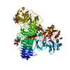
| ||||||||
|---|---|---|---|---|---|---|---|---|---|
| 1 |
| ||||||||
| Unit cell |
|
-Components
-Protein , 1 types, 1 molecules A
| #1: Protein | Mass: 145486.547 Da / Num. of mol.: 1 Source method: isolated from a genetically manipulated source Source: (gene. exp.) Homo sapiens (human) / Gene: CEMIP2, KIAA1412, TMEM2 / Plasmid: pCEP-Pu / Cell line (production host): Expi293 / Production host: Homo sapiens (human) / References: UniProt: Q9UHN6, hyaluronoglucosaminidase |
|---|
-Sugars , 6 types, 9 molecules 
| #2: Polysaccharide | Source method: isolated from a genetically manipulated source #3: Polysaccharide | Source method: isolated from a genetically manipulated source #4: Polysaccharide | beta-D-mannopyranose-(1-4)-2-acetamido-2-deoxy-beta-D-glucopyranose-(1-4)-2-acetamido-2-deoxy-beta- ...beta-D-mannopyranose-(1-4)-2-acetamido-2-deoxy-beta-D-glucopyranose-(1-4)-2-acetamido-2-deoxy-beta-D-glucopyranose | Source method: isolated from a genetically manipulated source #5: Polysaccharide | alpha-D-mannopyranose-(1-6)-beta-D-mannopyranose-(1-4)-2-acetamido-2-deoxy-beta-D-glucopyranose-(1- ...alpha-D-mannopyranose-(1-6)-beta-D-mannopyranose-(1-4)-2-acetamido-2-deoxy-beta-D-glucopyranose-(1-4)-[alpha-L-fucopyranose-(1-6)]2-acetamido-2-deoxy-beta-D-glucopyranose | Source method: isolated from a genetically manipulated source #6: Polysaccharide | alpha-D-mannopyranose-(1-3)-beta-D-mannopyranose-(1-4)-2-acetamido-2-deoxy-beta-D-glucopyranose-(1- ...alpha-D-mannopyranose-(1-3)-beta-D-mannopyranose-(1-4)-2-acetamido-2-deoxy-beta-D-glucopyranose-(1-4)-2-acetamido-2-deoxy-beta-D-glucopyranose | Source method: isolated from a genetically manipulated source #9: Sugar |  Type: D-saccharide, beta linking / Mass: 221.208 Da / Num. of mol.: 2 / Source method: obtained synthetically / Formula: C8H15NO6 Type: D-saccharide, beta linking / Mass: 221.208 Da / Num. of mol.: 2 / Source method: obtained synthetically / Formula: C8H15NO6 |
|---|
-Non-polymers , 2 types, 2 molecules 

| #7: Chemical | ChemComp-NI /  Mass: 58.693 Da / Num. of mol.: 1 / Source method: obtained synthetically / Formula: Ni Mass: 58.693 Da / Num. of mol.: 1 / Source method: obtained synthetically / Formula: Ni |
|---|---|
| #8: Chemical | ChemComp-CL /  Mass: 35.453 Da / Num. of mol.: 1 / Source method: obtained synthetically / Formula: Cl Mass: 35.453 Da / Num. of mol.: 1 / Source method: obtained synthetically / Formula: Cl |
-Details
| Has ligand of interest | N |
|---|---|
| Has protein modification | Y |
-Experimental details
-Experiment
| Experiment | Method: X-RAY DIFFRACTION / Number of used crystals: 1 |
|---|
- Sample preparation
| Crystal | Density Matthews: 3.09 Å3/Da / Density % sol: 60.21 % |
|---|---|
| Crystal grow | Temperature: 293 K / Method: vapor diffusion, sitting drop / pH: 4.5 Details: 0.1 M sodium acetate, pH 4.5, 30% polyethyleneglycol 3000 |
-Data collection
| Diffraction | Mean temperature: 100 K / Serial crystal experiment: N |
|---|---|
| Diffraction source | Source: SYNCHROTRON / Site: Diamond / Beamline: I04 / Wavelength: 0.9795 Å |
| Detector | Type: DECTRIS EIGER2 S 16M / Detector: PIXEL / Date: Feb 3, 2022 |
| Radiation | Protocol: SINGLE WAVELENGTH / Monochromatic (M) / Laue (L): M / Scattering type: x-ray |
| Radiation wavelength | Wavelength: 0.9795 Å / Relative weight: 1 |
| Reflection | Resolution: 3.5→92.35 Å / Num. obs: 23570 / % possible obs: 100 % / Redundancy: 26.8 % / CC1/2: 0.997 / Rpim(I) all: 0.073 / Net I/σ(I): 7.1 |
| Reflection shell | Resolution: 3.5→3.56 Å / Mean I/σ(I) obs: 0.5 / Num. unique obs: 1159 / CC1/2: 0.424 / Rpim(I) all: 0.871 |
- Processing
| Software |
| |||||||||||||||||||||||||||||||||||||||||||||||||||||||||||||||
|---|---|---|---|---|---|---|---|---|---|---|---|---|---|---|---|---|---|---|---|---|---|---|---|---|---|---|---|---|---|---|---|---|---|---|---|---|---|---|---|---|---|---|---|---|---|---|---|---|---|---|---|---|---|---|---|---|---|---|---|---|---|---|---|---|
| Refinement | Method to determine structure: MOLECULAR REPLACEMENT / Resolution: 3.5→91.55 Å / SU ML: 0.66 / Cross valid method: FREE R-VALUE / σ(F): 1.34 / Phase error: 34.92 / Stereochemistry target values: ML
| |||||||||||||||||||||||||||||||||||||||||||||||||||||||||||||||
| Solvent computation | Shrinkage radii: 0.9 Å / VDW probe radii: 1.11 Å / Solvent model: FLAT BULK SOLVENT MODEL | |||||||||||||||||||||||||||||||||||||||||||||||||||||||||||||||
| Refinement step | Cycle: LAST / Resolution: 3.5→91.55 Å
| |||||||||||||||||||||||||||||||||||||||||||||||||||||||||||||||
| Refine LS restraints |
| |||||||||||||||||||||||||||||||||||||||||||||||||||||||||||||||
| LS refinement shell |
|
