 Movie
Movie Controller

[English] 日本語
 Yorodumi
- PDB-8c6e: CRYSTAL STRUCTURE OF ODORANT BINDING PROTEIN 4 FROM ANOPHELES GAM... -
+ Open data

- Basic information
| Entry | Database: PDB / ID: 8c6e | |||||||||
|---|---|---|---|---|---|---|---|---|---|---|
| Title | CRYSTAL STRUCTURE OF ODORANT BINDING PROTEIN 4 FROM ANOPHELES GAMBIAE (AGAMOBP4) AT PH 8.5 | |||||||||
 Components Components | AGAP010489-PA | |||||||||
 Keywords Keywords | TRANSPORT PROTEIN / Odorant Binding Protein (OBP) / mosquito / olfaction | |||||||||
| Function / homology |  Function and homology information Function and homology informationdibutyl phthalate binding / olfactory behavior / odorant binding / sensory perception of smell / extracellular region / metal ion binding Similarity search - Function | |||||||||
| Biological species |  | |||||||||
| Method |  X-RAY DIFFRACTION / SYNCHROTRON / MOLECULAR REPLACEMENT / Resolution: 2.05 Å X-RAY DIFFRACTION / SYNCHROTRON / MOLECULAR REPLACEMENT / Resolution: 2.05 Å | |||||||||
 Authors Authors | Tsitsanou, K.E. / Drakou, C.E. / Zographos, S.E. | |||||||||
| Funding support |  Greece, 2items Greece, 2items
| |||||||||
 Citation Citation | Journal: Int.J.Biol.Macromol. / Year: 2023 Title: Influence of pH on indole-dependent heterodimeric interactions between Anopheles gambiae odorant-binding proteins OBP1 and OBP4. Authors: Mam, B. / Tsitsanou, K.E. / Liggri, P.G.V. / Saitta, F. / Stamati, E.C.V. / Mahita, J. / Leonis, G. / Drakou, C.E. / Papadopoulos, M. / Arnaud, P. / Offmann, B. / Fessas, D. / Sowdhamini, R. / Zographos, S.E. | |||||||||
| History |
|
- Structure visualization
| Structure viewer | Molecule: MolmilJmol/JSmol |
|---|
- Downloads & links
-Download
| PDBx/mmCIF format | 8c6e.cif.gz | 68.1 KB | Display | PDBx/mmCIF format |
|---|---|---|---|---|
| PDB format | pdb8c6e.ent.gz | 49.2 KB | Display | PDB format |
| PDBx/mmJSON format | 8c6e.json.gz | Tree view | PDBx/mmJSON format | |
| Others |  Other downloads Other downloads |
-Validation report
| Summary document | 8c6e_validation.pdf.gz | 414.7 KB | Display | wwPDB validaton report |
|---|---|---|---|---|
| Full document | 8c6e_full_validation.pdf.gz | 416.1 KB | Display | |
| Data in XML | 8c6e_validation.xml.gz | 7.7 KB | Display | |
| Data in CIF | 8c6e_validation.cif.gz | 9.7 KB | Display | |
| Arichive directory | https://data.pdbj.org/pub/pdb/validation_reports/c6/8c6eftp://data.pdbj.org/pub/pdb/validation_reports/c6/8c6e | HTTPS FTP |
-Related structure data
| Related structure data | 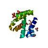 8c68C  3n88 C: citing same article ( S: Starting model for refinement |
|---|---|
| Similar structure data |
-Links
 PDBj
PDBj


- Assembly
| Deposited unit | 
| ||||||||
|---|---|---|---|---|---|---|---|---|---|
| 1 |
| ||||||||
| Unit cell |
|
-Components
| #1: Protein | Mass: 14048.541 Da / Num. of mol.: 1 Source method: isolated from a genetically manipulated source Source: (gene. exp.) Gene: OBP-4, 1275228, AgamOBP4, OBP4, AgaP_AGAP010489, agCG48601 Plasmid: PET-22B(+) / Production host:  |
|---|---|
| #2: Chemical | ChemComp-FE2 /   Mass: 55.845 Da / Num. of mol.: 1 / Source method: obtained synthetically / Formula: Fe Mass: 55.845 Da / Num. of mol.: 1 / Source method: obtained synthetically / Formula: Fe |
| #3: Chemical | ChemComp-MG /   Mass: 24.305 Da / Num. of mol.: 1 / Source method: obtained synthetically / Formula: Mg Mass: 24.305 Da / Num. of mol.: 1 / Source method: obtained synthetically / Formula: Mg |
| #4: Water | ChemComp-HOH /  Mass: 18.015 Da / Num. of mol.: 70 / Source method: isolated from a natural source / Formula: H2O Mass: 18.015 Da / Num. of mol.: 70 / Source method: isolated from a natural source / Formula: H2O |
| Has ligand of interest | N |
| Has protein modification | Y |
-Experimental details
-Experiment
| Experiment | Method: X-RAY DIFFRACTION / Number of used crystals: 1 |
|---|
- Sample preparation
| Crystal | Density Matthews: 1.89 Å3/Da / Density % sol: 34.89 % |
|---|---|
| Crystal grow | Temperature: 289 K / Method: vapor diffusion, sitting drop / pH: 8.5 Details: 20% W/V PEG 8000, 0.2 M MAGNESIUM CHLORIDE, 0.1 M TRIS-HCL PH 8.5, |
-Data collection
| Diffraction | Mean temperature: 100 K / Serial crystal experiment: N |
|---|---|
| Diffraction source | Source: SYNCHROTRON / Site: MAX II  / Beamline: I911-2 / Wavelength: 1.03796 Å / Beamline: I911-2 / Wavelength: 1.03796 Å |
| Detector | Type: MAR CCD 165 mm / Detector: CCD / Date: Nov 13, 2009 / Details: MULTILAYER MIRROR |
| Radiation | Protocol: SINGLE WAVELENGTH / Monochromatic (M) / Laue (L): M / Scattering type: x-ray |
| Radiation wavelength | Wavelength: 1.03796 Å / Relative weight: 1 |
| Reflection | Resolution: 2.05→39.54 Å / Num. obs: 7086 / % possible obs: 99.8 % / Redundancy: 7 % / Rpim(I) all: 0.025 / Rrim(I) all: 0.067 / Net I/σ(I): 21.2 |
| Reflection shell | Resolution: 2.05→2.16 Å / % possible obs: 100 % / Redundancy: 7.1 % / Num. measured all: 7131 / Num. unique obs: 1001 / Rpim(I) all: 0.067 / Rrim(I) all: 0.183 / Net I/σ(I) obs: 9.8 |
- Processing
| Software |
| ||||||||||||||||||||||||||||||||||||||||||||||||||||||||||||||||||||||||||||||||||||||||||||||||||||||||||||||||||||||||||||||||||||||||||||||||||||||||||||||||||||||||||||||||||||||
|---|---|---|---|---|---|---|---|---|---|---|---|---|---|---|---|---|---|---|---|---|---|---|---|---|---|---|---|---|---|---|---|---|---|---|---|---|---|---|---|---|---|---|---|---|---|---|---|---|---|---|---|---|---|---|---|---|---|---|---|---|---|---|---|---|---|---|---|---|---|---|---|---|---|---|---|---|---|---|---|---|---|---|---|---|---|---|---|---|---|---|---|---|---|---|---|---|---|---|---|---|---|---|---|---|---|---|---|---|---|---|---|---|---|---|---|---|---|---|---|---|---|---|---|---|---|---|---|---|---|---|---|---|---|---|---|---|---|---|---|---|---|---|---|---|---|---|---|---|---|---|---|---|---|---|---|---|---|---|---|---|---|---|---|---|---|---|---|---|---|---|---|---|---|---|---|---|---|---|---|---|---|---|---|
| Refinement | Method to determine structure: MOLECULAR REPLACEMENT Starting model: PDB ENTRY 3N88 3n88 Resolution: 2.05→39.54 Å / Cor.coef. Fo:Fc: 0.942 / Cor.coef. Fo:Fc free: 0.927 / SU B: 9.383 / SU ML: 0.132 / Cross valid method: THROUGHOUT / σ(F): 0 / ESU R: 0.262 / ESU R Free: 0.194 / Stereochemistry target values: MAXIMUM LIKELIHOOD / Details: HYDROGENS HAVE BEEN ADDED IN THE RIDING POSITIONS
| ||||||||||||||||||||||||||||||||||||||||||||||||||||||||||||||||||||||||||||||||||||||||||||||||||||||||||||||||||||||||||||||||||||||||||||||||||||||||||||||||||||||||||||||||||||||
| Solvent computation | Ion probe radii: 0.8 Å / Shrinkage radii: 0.8 Å / VDW probe radii: 1.4 Å / Solvent model: MASK | ||||||||||||||||||||||||||||||||||||||||||||||||||||||||||||||||||||||||||||||||||||||||||||||||||||||||||||||||||||||||||||||||||||||||||||||||||||||||||||||||||||||||||||||||||||||
| Displacement parameters | Biso mean: 23.63 Å2
| ||||||||||||||||||||||||||||||||||||||||||||||||||||||||||||||||||||||||||||||||||||||||||||||||||||||||||||||||||||||||||||||||||||||||||||||||||||||||||||||||||||||||||||||||||||||
| Refinement step | Cycle: LAST / Resolution: 2.05→39.54 Å
| ||||||||||||||||||||||||||||||||||||||||||||||||||||||||||||||||||||||||||||||||||||||||||||||||||||||||||||||||||||||||||||||||||||||||||||||||||||||||||||||||||||||||||||||||||||||
| Refine LS restraints |
|