 Movie
Movie Controller


+ Open data

- Basic information
| Entry | Database: PDB / ID: 8c5r | |||||||||||||||||||||||||||||||||||||||
|---|---|---|---|---|---|---|---|---|---|---|---|---|---|---|---|---|---|---|---|---|---|---|---|---|---|---|---|---|---|---|---|---|---|---|---|---|---|---|---|---|
| Title | Omicron B.1.1.529 2 RBD up conformation | |||||||||||||||||||||||||||||||||||||||
 Components Components | Spike glycoprotein | |||||||||||||||||||||||||||||||||||||||
 Keywords Keywords | VIRAL PROTEIN / COVID-19 / SPIKE TRIMER / OMICRON / B.1.1.529 | |||||||||||||||||||||||||||||||||||||||
| Function / homology |  Function and homology information Function and homology informationpositive regulation of viral entry into host cell / host cell endoplasmic reticulum-Golgi intermediate compartment membrane / receptor-mediated virion attachment to host cell / host cell surface receptor binding / endocytosis involved in viral entry into host cell / fusion of virus membrane with host plasma membrane / fusion of virus membrane with host endosome membrane / viral envelope / host cell plasma membrane / virion membrane ...positive regulation of viral entry into host cell / host cell endoplasmic reticulum-Golgi intermediate compartment membrane / receptor-mediated virion attachment to host cell / host cell surface receptor binding / endocytosis involved in viral entry into host cell / fusion of virus membrane with host plasma membrane / fusion of virus membrane with host endosome membrane / viral envelope / host cell plasma membrane / virion membrane / membrane / identical protein binding Similarity search - Function | |||||||||||||||||||||||||||||||||||||||
| Biological species |  Alphacoronavirus Alphacoronavirus | |||||||||||||||||||||||||||||||||||||||
| Method | ELECTRON MICROSCOPY / single particle reconstruction / cryo EM / Resolution: 3.7 Å | |||||||||||||||||||||||||||||||||||||||
 Authors Authors | Raghavan, S.S.R. / Walker, M.R. / Salanti, A. / Barfod, L.K. / Wang, K.T. | |||||||||||||||||||||||||||||||||||||||
| Funding support |  Denmark, 3items Denmark, 3items
| |||||||||||||||||||||||||||||||||||||||
 Citation Citation | Journal: Commun Biol / Year: 2024 Title: Broadly potent spike-specific human monoclonal antibodies inhibit SARS-CoV-2 Omicron sub-lineages. Authors: Melanie R Walker / Alexander Underwood / Kasper H Björnsson / Sai Sundar Rajan Raghavan / Maria R Bassi / Alekxander Binderup / Long V Pham / Santseharay Ramirez / Mette Pinholt / Robert ...Authors: Melanie R Walker / Alexander Underwood / Kasper H Björnsson / Sai Sundar Rajan Raghavan / Maria R Bassi / Alekxander Binderup / Long V Pham / Santseharay Ramirez / Mette Pinholt / Robert Dagil / Anne S Knudsen / Manja Idorn / Max Soegaard / Kaituo Wang / Andrew B Ward / Ali Salanti / Jens Bukh / Lea Barfod /  Abstract: The continuous emergence of SARS-CoV-2 variants of concern has rendered many therapeutic monoclonal antibodies (mAbs) ineffective. To date, there are no clinically authorized therapeutic antibodies ...The continuous emergence of SARS-CoV-2 variants of concern has rendered many therapeutic monoclonal antibodies (mAbs) ineffective. To date, there are no clinically authorized therapeutic antibodies effective against the recently circulating Omicron sub-lineages BA.2.86 and JN.1. Here, we report the isolation of broad and potent neutralizing human mAbs (HuMabs) from a healthcare worker infected with SARS-CoV-2 early in the pandemic. These include a genetically unique HuMab, named K501SP6, which can neutralize different Omicron sub-lineages, including BQ.1, XBB.1, BA.2.86 and JN.1, by targeting a highly conserved epitope on the N terminal domain, as well as an RBD-specific HuMab (K501SP3) with high potency towards earlier circulating variants that was escaped by the more recent Omicron sub-lineages through spike F486 and E484 substitutions. Characterizing SARS-CoV-2 spike-specific HuMabs, including broadly reactive non-RBD-specific HuMabs, can give insight into the immune mechanisms involved in neutralization and immune evasion, which can be a valuable addition to already existing SARS-CoV-2 therapies. | |||||||||||||||||||||||||||||||||||||||
| History |
|
- Structure visualization
| Structure viewer | Molecule: MolmilJmol/JSmol |
|---|
- Downloads & links
-Download
| PDBx/mmCIF format | 8c5r.cif.gz | 549.6 KB | Display | PDBx/mmCIF format |
|---|---|---|---|---|
| PDB format | pdb8c5r.ent.gz | 424.8 KB | Display | PDB format |
| PDBx/mmJSON format | 8c5r.json.gz | Tree view | PDBx/mmJSON format | |
| Others |  Other downloads Other downloads |
-Validation report
| Arichive directory | https://data.pdbj.org/pub/pdb/validation_reports/c5/8c5rftp://data.pdbj.org/pub/pdb/validation_reports/c5/8c5r | HTTPS FTP |
|---|
-Related structure data
| Related structure data |  16441MC 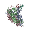 9fjkC M: map data used to model this data C: citing same article ( |
|---|---|
| Similar structure data |
-Links
 PDBj
PDBj- Assembly
| Deposited unit | 
|
|---|---|
| 1 |
|
-Components
| #1: Protein | Mass: 118193.594 Da / Num. of mol.: 3 Source method: isolated from a genetically manipulated source Source: (gene. exp.) Alphacoronavirus / Production host:  Homo sapiens (human) / References: UniProt: A0A6M4AIH4 Homo sapiens (human) / References: UniProt: A0A6M4AIH4Has protein modification | Y | |
|---|
-Experimental details
-Experiment
| Experiment | Method: ELECTRON MICROSCOPY |
|---|---|
| EM experiment | Aggregation state: PARTICLE / 3D reconstruction method: single particle reconstruction |
- Sample preparation
| Component | Name: Omicron Spike trimeric complex / Type: COMPLEX / Entity ID: all / Source: RECOMBINANT |
|---|---|
| Molecular weight | Value: 0.4 kDa/nm / Experimental value: YES |
| Source (natural) | Organism: Alphacoronavirus |
| Source (recombinant) | Organism: Homo sapiens (human) |
| Buffer solution | pH: 7.5 |
| Specimen | Conc.: 0.1 mg/ml / Embedding applied: NO / Shadowing applied: NO / Staining applied: NO / Vitrification applied: YES |
| Specimen support | Grid material: GRAPHENE OXIDE / Grid type: Homemade |
| Vitrification | Cryogen name: ETHANE |
- Electron microscopy imaging
| Experimental equipment |  Model: Titan Krios / Image courtesy: FEI Company |
|---|---|
| Microscopy | Model: FEI TITAN KRIOS |
| Electron gun | Electron source:  FIELD EMISSION GUN / Accelerating voltage: 300 kV / Illumination mode: FLOOD BEAM FIELD EMISSION GUN / Accelerating voltage: 300 kV / Illumination mode: FLOOD BEAM |
| Electron lens | Mode: BRIGHT FIELD / Nominal defocus max: 2900 nm / Nominal defocus min: 1000 nm |
| Image recording | Electron dose: 42 e/Å2 / Detector mode: COUNTING / Film or detector model: FEI FALCON III (4k x 4k) |
- Processing
| Software | Name: PHENIX / Version: 1.19.2_4158: / Classification: refinement |
|---|---|
| EM software | Name: PHENIX / Category: model refinement |
| CTF correction | Type: NONE |
| 3D reconstruction | Resolution: 3.7 Å / Resolution method: FSC 0.143 CUT-OFF / Num. of particles: 19500 / Symmetry type: POINT |
| Atomic model building | Protocol: RIGID BODY FIT |
