 Movie
Movie Controller

[English] 日本語
 Yorodumi
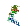
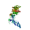
- PDB-8c5b: Crystal Structure of Penicillin-binding Protein 3 (PBP3) from Sta... -
+ Open data

- Basic information
| Entry | Database: PDB / ID: 8c5b | ||||||
|---|---|---|---|---|---|---|---|
| Title | Crystal Structure of Penicillin-binding Protein 3 (PBP3) from Staphylococcus Epidermidis | ||||||
 Components Components | Penicillin-binding protein 3 | ||||||
 Keywords Keywords | PENICILLIN-BINDING PROTEIN | ||||||
| Function / homology |  Function and homology information Function and homology informationpeptidoglycan L,D-transpeptidase activity / penicillin binding / peptidoglycan biosynthetic process / cell wall organization / regulation of cell shape / plasma membrane Similarity search - Function | ||||||
| Biological species |  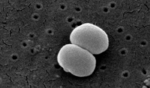 Staphylococcus epidermidis (bacteria) Staphylococcus epidermidis (bacteria) | ||||||
| Method |  X-RAY DIFFRACTION / SYNCHROTRON / MOLECULAR REPLACEMENT / Resolution: 2.5 Å X-RAY DIFFRACTION / SYNCHROTRON / MOLECULAR REPLACEMENT / Resolution: 2.5 Å | ||||||
 Authors Authors | Schwinzer, M. / Brognaro, H. / Rohde, H. / Betzel, C. | ||||||
| Funding support |  Germany, 1items Germany, 1items
| ||||||
 Citation Citation | Journal: Int J Appl Biol Pharm / Year: 2023 Title: Structure and Dynamics of the Penicillin-Binding Protein 3 from Staphylococcus Epidermidis Native and in Complex with Cefotaxime and Vaborbactam Authors: Schwinzer, M. / Brognaro, H. / Rohde, H. / Betzel, C. | ||||||
| History |
|
- Structure visualization
| Structure viewer | Molecule: MolmilJmol/JSmol |
|---|
- Downloads & links
-Download
| PDBx/mmCIF format | 8c5b.cif.gz | 304.3 KB | Display | PDBx/mmCIF format |
|---|---|---|---|---|
| PDB format | pdb8c5b.ent.gz | 205.3 KB | Display | PDB format |
| PDBx/mmJSON format | 8c5b.json.gz | Tree view | PDBx/mmJSON format | |
| Others |  Other downloads Other downloads |
-Validation report
| Arichive directory | https://data.pdbj.org/pub/pdb/validation_reports/c5/8c5bftp://data.pdbj.org/pub/pdb/validation_reports/c5/8c5b | HTTPS FTP |
|---|
-Related structure data
-Links
 PDBj
PDBj
- Assembly
| Deposited unit | 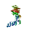
| ||||||||||||
|---|---|---|---|---|---|---|---|---|---|---|---|---|---|
| 1 |
| ||||||||||||
| Unit cell |
|
-Components
| #1: Protein | Mass: 74932.000 Da / Num. of mol.: 1 Source method: isolated from a genetically manipulated source Source: (gene. exp.) Staphylococcus epidermidis (strain ATCC 35984 / RP62A) (bacteria)Gene: pbp3, SERP1117 / Production host: |
|---|
-Experimental details
-Experiment
| Experiment | Method: X-RAY DIFFRACTION / Number of used crystals: 1 |
|---|
- Sample preparation
| Crystal | Density Matthews: 3.57 Å3/Da / Density % sol: 65.52 % |
|---|---|
| Crystal grow | Temperature: 293.15 K / Method: vapor diffusion, hanging drop / Details: Ammonium sulfate |
-Data collection
| Diffraction | Mean temperature: 100 K / Serial crystal experiment: N |
|---|---|
| Diffraction source | Source: SYNCHROTRON / Site: PETRA III, EMBL c/o DESY / Beamline: P13 (MX1) / Wavelength: 0.97625 Å |
| Detector | Type: DECTRIS EIGER X 16M / Detector: PIXEL / Date: Jun 24, 2021 |
| Radiation | Protocol: SINGLE WAVELENGTH / Monochromatic (M) / Laue (L): M / Scattering type: x-ray |
| Radiation wavelength | Wavelength: 0.97625 Å / Relative weight: 1 |
| Reflection | Resolution: 2.067→80.687 Å / Num. obs: 46353 / % possible obs: 95.2 % / Redundancy: 18.8 % / Biso Wilson estimate: 56.24 Å2 / CC1/2: 0.999 / Net I/σ(I): 16.8 |
| Reflection shell | Resolution: 2.067→2.297 Å / Num. unique obs: 2318 / CC1/2: 0.996 |
- Processing
| Software |
| ||||||||||||||||||||||||||||||||||||||||||||||||||||||||||||||||||||||||||||||||||||||||||||||||||
|---|---|---|---|---|---|---|---|---|---|---|---|---|---|---|---|---|---|---|---|---|---|---|---|---|---|---|---|---|---|---|---|---|---|---|---|---|---|---|---|---|---|---|---|---|---|---|---|---|---|---|---|---|---|---|---|---|---|---|---|---|---|---|---|---|---|---|---|---|---|---|---|---|---|---|---|---|---|---|---|---|---|---|---|---|---|---|---|---|---|---|---|---|---|---|---|---|---|---|---|
| Refinement | Method to determine structure: MOLECULAR REPLACEMENT / Resolution: 2.5→57.94 Å / SU ML: 0.3166 / Cross valid method: FREE R-VALUE / σ(F): 1.33 / Phase error: 26.7949 Stereochemistry target values: GeoStd + Monomer Library + CDL v1.2
| ||||||||||||||||||||||||||||||||||||||||||||||||||||||||||||||||||||||||||||||||||||||||||||||||||
| Solvent computation | Shrinkage radii: 0.9 Å / VDW probe radii: 1.1 Å / Solvent model: FLAT BULK SOLVENT MODEL | ||||||||||||||||||||||||||||||||||||||||||||||||||||||||||||||||||||||||||||||||||||||||||||||||||
| Displacement parameters | Biso mean: 71.95 Å2 | ||||||||||||||||||||||||||||||||||||||||||||||||||||||||||||||||||||||||||||||||||||||||||||||||||
| Refinement step | Cycle: LAST / Resolution: 2.5→57.94 Å
| ||||||||||||||||||||||||||||||||||||||||||||||||||||||||||||||||||||||||||||||||||||||||||||||||||
| Refine LS restraints |
| ||||||||||||||||||||||||||||||||||||||||||||||||||||||||||||||||||||||||||||||||||||||||||||||||||
| LS refinement shell |
| ||||||||||||||||||||||||||||||||||||||||||||||||||||||||||||||||||||||||||||||||||||||||||||||||||
| Refinement TLS params. | Method: refined / Refine-ID: X-RAY DIFFRACTION
| ||||||||||||||||||||||||||||||||||||||||||||||||||||||||||||||||||||||||||||||||||||||||||||||||||
| Refinement TLS group | Refine-ID: X-RAY DIFFRACTION / Auth asym-ID: A / Label asym-ID: A
|
