 Movie
Movie Controller


+ Open data

- Basic information
| Entry | Database: PDB / ID: 8c3o | ||||||
|---|---|---|---|---|---|---|---|
| Title | Crystal structure of autotaxin gamma and compound MEY-003 | ||||||
 Components Components | Ectonucleotide pyrophosphatase/phosphodiesterase family member 2 | ||||||
 Keywords Keywords | HYDROLASE / Inhibitor / co-crystal / ectonucleotide pyrophosphatase/phosphodiesterase (ENPP) | ||||||
| Function / homology |  Function and homology information Function and homology informationVitamin B5 (pantothenate) metabolism / alkylglycerophosphoethanolamine phosphodiesterase / sphingolipid catabolic process / phospholipase D / phosphatidylcholine catabolic process / phospholipid catabolic process / D-type glycerophospholipase activity / positive regulation of lamellipodium morphogenesis / phosphodiesterase I activity / phosphatidylcholine lysophospholipase A1 activity ...Vitamin B5 (pantothenate) metabolism / alkylglycerophosphoethanolamine phosphodiesterase / sphingolipid catabolic process / phospholipase D / phosphatidylcholine catabolic process / phospholipid catabolic process / D-type glycerophospholipase activity / positive regulation of lamellipodium morphogenesis / phosphodiesterase I activity / phosphatidylcholine lysophospholipase A1 activity / scavenger receptor activity / alkylglycerophosphoethanolamine phosphodiesterase activity / polysaccharide binding / positive regulation of epithelial cell migration / regulation of cell migration / chemotaxis / nucleic acid binding / immune response / hydrolase activity / calcium ion binding / : / zinc ion binding / plasma membrane Similarity search - Function | ||||||
| Biological species |  Homo sapiens (human) Homo sapiens (human) | ||||||
| Method |  X-RAY DIFFRACTION / SYNCHROTRON / MOLECULAR REPLACEMENT / Resolution: 2.47 Å X-RAY DIFFRACTION / SYNCHROTRON / MOLECULAR REPLACEMENT / Resolution: 2.47 Å | ||||||
 Authors Authors | Eymery, M.C. / McCarthy, A.A. | ||||||
| Funding support | European Union, 1items
| ||||||
 Citation Citation | Journal: Eur.J.Med.Chem. / Year: 2023 Title: Discovery of potent chromone-based autotaxin inhibitors inspired by cannabinoids. Authors: Eymery, M.C. / Nguyen, K.A. / Basu, S. / Hausmann, J. / Tran-Nguyen, V.K. / Seidel, H.P. / Gutierrez, L. / Boumendjel, A. / McCarthy, A.A. #1: Journal: Acta Crystallogr D Biol Crystallogr / Year: 2012 Title: Towards automated crystallographic structure refinement with phenix.refine. Authors: Afonine, P.V. / Grosse-Kunstleve, R.W. / Echols, N. / Headd, J.J. / Moriarty, N.W. / Mustyakimov, M. / Terwilliger, T.C. / Urzhumtsev, A. / Zwart, P.H. / Adams, P.D. #2: Journal: Acta Crystallogr D Struct Biol / Year: 2019 Title: Macromolecular structure determination using X-rays, neutrons and electrons: recent developments in Phenix. Authors: Dorothee Liebschner / Pavel V Afonine / Matthew L Baker / Gábor Bunkóczi / Vincent B Chen / Tristan I Croll / Bradley Hintze / Li Wei Hung / Swati Jain / Airlie J McCoy / Nigel W Moriarty ...Authors: Dorothee Liebschner / Pavel V Afonine / Matthew L Baker / Gábor Bunkóczi / Vincent B Chen / Tristan I Croll / Bradley Hintze / Li Wei Hung / Swati Jain / Airlie J McCoy / Nigel W Moriarty / Robert D Oeffner / Billy K Poon / Michael G Prisant / Randy J Read / Jane S Richardson / David C Richardson / Massimo D Sammito / Oleg V Sobolev / Duncan H Stockwell / Thomas C Terwilliger / Alexandre G Urzhumtsev / Lizbeth L Videau / Christopher J Williams / Paul D Adams /    Abstract: Diffraction (X-ray, neutron and electron) and electron cryo-microscopy are powerful methods to determine three-dimensional macromolecular structures, which are required to understand biological ...Diffraction (X-ray, neutron and electron) and electron cryo-microscopy are powerful methods to determine three-dimensional macromolecular structures, which are required to understand biological processes and to develop new therapeutics against diseases. The overall structure-solution workflow is similar for these techniques, but nuances exist because the properties of the reduced experimental data are different. Software tools for structure determination should therefore be tailored for each method. Phenix is a comprehensive software package for macromolecular structure determination that handles data from any of these techniques. Tasks performed with Phenix include data-quality assessment, map improvement, model building, the validation/rebuilding/refinement cycle and deposition. Each tool caters to the type of experimental data. The design of Phenix emphasizes the automation of procedures, where possible, to minimize repetitive and time-consuming manual tasks, while default parameters are chosen to encourage best practice. A graphical user interface provides access to many command-line features of Phenix and streamlines the transition between programs, project tracking and re-running of previous tasks. | ||||||
| History |
|
- Structure visualization
| Structure viewer | Molecule: MolmilJmol/JSmol |
|---|
- Downloads & links
-Download
| PDBx/mmCIF format | 8c3o.cif.gz | 768.7 KB | Display | PDBx/mmCIF format |
|---|---|---|---|---|
| PDB format | pdb8c3o.ent.gz | 520.2 KB | Display | PDB format |
| PDBx/mmJSON format | 8c3o.json.gz | Tree view | PDBx/mmJSON format | |
| Others |  Other downloads Other downloads |
-Validation report
| Arichive directory | https://data.pdbj.org/pub/pdb/validation_reports/c3/8c3oftp://data.pdbj.org/pub/pdb/validation_reports/c3/8c3o | HTTPS FTP |
|---|
-Related structure data
| Related structure data |  8c3pC 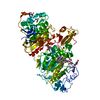 8c4wC 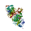 8c7rC 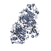 4zg6S S: Starting model for refinement C: citing same article ( |
|---|---|
| Similar structure data |
-Links
 PDBj
PDBj
- Assembly
| Deposited unit | 
| ||||||||||||
|---|---|---|---|---|---|---|---|---|---|---|---|---|---|
| 1 | 
| ||||||||||||
| 2 | 
| ||||||||||||
| Unit cell |
|
-Components
-Protein , 1 types, 2 molecules AB
| #1: Protein | Mass: 102023.867 Da / Num. of mol.: 2 / Mutation: N54A, N411A Source method: isolated from a genetically manipulated source Source: (gene. exp.) Homo sapiens (human) / Gene: ENPP2, ATX, PDNP2 / Plasmid: pcDNA5.1 / Cell line (production host): HEK293 / Organ (production host): Kidney / Production host: Homo sapiens (human)References: UniProt: Q13822, alkylglycerophosphoethanolamine phosphodiesterase |
|---|
-Sugars , 2 types, 2 molecules
| #2: Polysaccharide | beta-D-mannopyranose-(1-4)-2-acetamido-2-deoxy-beta-D-glucopyranose-(1-4)-2-acetamido-2-deoxy-beta- ...beta-D-mannopyranose-(1-4)-2-acetamido-2-deoxy-beta-D-glucopyranose-(1-4)-2-acetamido-2-deoxy-beta-D-glucopyranose Source method: isolated from a genetically manipulated source |
|---|---|
| #3: Polysaccharide | 2-acetamido-2-deoxy-beta-D-glucopyranose-(1-4)-2-acetamido-2-deoxy-beta-D-glucopyranose Source method: isolated from a genetically manipulated source |
-Non-polymers , 7 types, 101 molecules 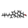
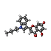


| #4: Chemical |  Mass: 402.653 Da / Num. of mol.: 2 / Source method: obtained synthetically / Formula: C27H46O2 Mass: 402.653 Da / Num. of mol.: 2 / Source method: obtained synthetically / Formula: C27H46O2#5: Chemical |  Mass: 363.406 Da / Num. of mol.: 2 / Source method: obtained synthetically / Formula: C22H21NO4 / Feature type: SUBJECT OF INVESTIGATION Mass: 363.406 Da / Num. of mol.: 2 / Source method: obtained synthetically / Formula: C22H21NO4 / Feature type: SUBJECT OF INVESTIGATION#6: Chemical | ChemComp-GOL /  Mass: 92.094 Da / Num. of mol.: 5 / Source method: obtained synthetically / Formula: C3H8O3 Mass: 92.094 Da / Num. of mol.: 5 / Source method: obtained synthetically / Formula: C3H8O3#7: Chemical | ChemComp-ZN /  Mass: 65.409 Da / Num. of mol.: 4 / Source method: obtained synthetically / Formula: Zn Mass: 65.409 Da / Num. of mol.: 4 / Source method: obtained synthetically / Formula: Zn#8: Chemical | ChemComp-IOD /  Mass: 126.904 Da / Num. of mol.: 37 / Source method: obtained synthetically / Formula: I Mass: 126.904 Da / Num. of mol.: 37 / Source method: obtained synthetically / Formula: I#9: Chemical |  Mass: 40.078 Da / Num. of mol.: 2 / Source method: obtained synthetically / Formula: Ca Mass: 40.078 Da / Num. of mol.: 2 / Source method: obtained synthetically / Formula: Ca#10: Water | ChemComp-HOH / | Mass: 18.015 Da / Num. of mol.: 49 / Source method: isolated from a natural source / Formula: H2O |
|---|
-Details
| Has ligand of interest | Y |
|---|---|
| Has protein modification | Y |
-Experimental details
-Experiment
| Experiment | Method: X-RAY DIFFRACTION / Number of used crystals: 1 |
|---|
- Sample preparation
| Crystal | Density Matthews: 2.35 Å3/Da / Density % sol: 47.72 % |
|---|---|
| Crystal grow | Temperature: 293 K / Method: vapor diffusion, hanging drop / pH: 8 Details: 18-22% PEG3350, 0,1-0,4M Ammonium Iodide, 0,1-0.4M Sodium Thiocyanate |
-Data collection
| Diffraction | Mean temperature: 100 K / Serial crystal experiment: N |
|---|---|
| Diffraction source | Source: SYNCHROTRON / Site: ESRF / Beamline: ID30B / Wavelength: 0.97625 Å |
| Detector | Type: DECTRIS PILATUS 6M / Detector: PIXEL / Date: May 4, 2022 |
| Radiation | Monochromator: Si111 channel cut mono / Protocol: SINGLE WAVELENGTH / Monochromatic (M) / Laue (L): M / Scattering type: x-ray |
| Radiation wavelength | Wavelength: 0.97625 Å / Relative weight: 1 |
| Reflection | Resolution: 2.468→144.239 Å / Num. obs: 45039 / % possible obs: 98.9 % / Redundancy: 3.3 % / Biso Wilson estimate: 25.67 Å2 / CC1/2: 0.964 / Net I/σ(I): 3.6 |
| Reflection shell | Resolution: 2.468→2.758 Å / Num. unique obs: 2191 / CC1/2: 0.52 |
- Processing
| Software |
| |||||||||||||||||||||||||||||||||||||||||||||||||||||||||||||||||||||||||||||||||||||||||||||||||||||||||||||||||||||||
|---|---|---|---|---|---|---|---|---|---|---|---|---|---|---|---|---|---|---|---|---|---|---|---|---|---|---|---|---|---|---|---|---|---|---|---|---|---|---|---|---|---|---|---|---|---|---|---|---|---|---|---|---|---|---|---|---|---|---|---|---|---|---|---|---|---|---|---|---|---|---|---|---|---|---|---|---|---|---|---|---|---|---|---|---|---|---|---|---|---|---|---|---|---|---|---|---|---|---|---|---|---|---|---|---|---|---|---|---|---|---|---|---|---|---|---|---|---|---|---|---|
| Refinement | Method to determine structure: MOLECULAR REPLACEMENT Starting model: 4ZG6 Resolution: 2.47→57.64 Å / SU ML: 0.2825 / Cross valid method: FREE R-VALUE / σ(F): 1.36 / Phase error: 28.436 Stereochemistry target values: GeoStd + Monomer Library + CDL v1.2
| |||||||||||||||||||||||||||||||||||||||||||||||||||||||||||||||||||||||||||||||||||||||||||||||||||||||||||||||||||||||
| Solvent computation | Shrinkage radii: 0.9 Å / VDW probe radii: 1.1 Å / Solvent model: FLAT BULK SOLVENT MODEL | |||||||||||||||||||||||||||||||||||||||||||||||||||||||||||||||||||||||||||||||||||||||||||||||||||||||||||||||||||||||
| Displacement parameters | Biso mean: 28.34 Å2 | |||||||||||||||||||||||||||||||||||||||||||||||||||||||||||||||||||||||||||||||||||||||||||||||||||||||||||||||||||||||
| Refinement step | Cycle: LAST / Resolution: 2.47→57.64 Å
| |||||||||||||||||||||||||||||||||||||||||||||||||||||||||||||||||||||||||||||||||||||||||||||||||||||||||||||||||||||||
| Refine LS restraints |
| |||||||||||||||||||||||||||||||||||||||||||||||||||||||||||||||||||||||||||||||||||||||||||||||||||||||||||||||||||||||
| LS refinement shell |
| |||||||||||||||||||||||||||||||||||||||||||||||||||||||||||||||||||||||||||||||||||||||||||||||||||||||||||||||||||||||
| Refinement TLS params. | Method: refined / Origin x: 16.3361149245 Å / Origin y: -50.7255025913 Å / Origin z: 41.0752972265 Å
| |||||||||||||||||||||||||||||||||||||||||||||||||||||||||||||||||||||||||||||||||||||||||||||||||||||||||||||||||||||||
| Refinement TLS group | Selection details: all |
