[English] 日本語
Yorodumi
- PDB-8c3c: 14-3-3 sigma with Pin1 binding site pS72 and bound Fusicoccin A -

+
Open data


ID or keywords:

Loading...

-
Basic information

Entry
Database: PDB / ID: 8c3c
Title14-3-3 sigma with Pin1 binding site pS72 and bound Fusicoccin A
Components
  • 14-3-3 protein sigma
  • Peptidyl-prolyl cis-trans isomerase NIMA-interacting 1
Keywords
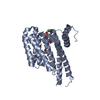
PEPTIDE BINDING PROTEIN / 14-3-3 sigma / Pin1 / stabilizer / Fusicoccin A
Function / homology
Function and homology information


cis-trans isomerase activity / phosphothreonine residue binding / negative regulation of cell motility / ubiquitin ligase activator activity / regulation of protein localization to nucleus / GTPase activating protein binding / postsynaptic cytosol / mitogen-activated protein kinase kinase binding / regulation of mitotic nuclear division / negative regulation of amyloid-beta formation ...cis-trans isomerase activity / phosphothreonine residue binding / negative regulation of cell motility / ubiquitin ligase activator activity / regulation of protein localization to nucleus / GTPase activating protein binding / postsynaptic cytosol / mitogen-activated protein kinase kinase binding / regulation of mitotic nuclear division / negative regulation of amyloid-beta formation / negative regulation of SMAD protein signal transduction / regulation of epidermal cell division / protein kinase C inhibitor activity / positive regulation of epidermal cell differentiation / keratinocyte development / keratinization / PI5P Regulates TP53 Acetylation / cytoskeletal motor activity / Regulation of localization of FOXO transcription factors / keratinocyte proliferation / RHO GTPases Activate NADPH Oxidases / phosphoserine residue binding / Activation of BAD and translocation to mitochondria / negative regulation of keratinocyte proliferation / establishment of skin barrier / SARS-CoV-2 targets host intracellular signalling and regulatory pathways / protein peptidyl-prolyl isomerization / Chk1/Chk2(Cds1) mediated inactivation of Cyclin B:Cdk1 complex / protein kinase A signaling / negative regulation of stem cell proliferation / SARS-CoV-1 targets host intracellular signalling and regulatory pathways / positive regulation of protein dephosphorylation / RHO GTPases activate PKNs / protein export from nucleus / negative regulation of innate immune response / protein sequestering activity / TP53 Regulates Transcription of Genes Involved in G2 Cell Cycle Arrest / release of cytochrome c from mitochondria / positive regulation of protein export from nucleus / ciliary basal body / negative regulation of protein binding / regulation of cytokinesis / peptidylprolyl isomerase / peptidyl-prolyl cis-trans isomerase activity / stem cell proliferation / Negative regulators of DDX58/IFIH1 signaling / Translocation of SLC2A4 (GLUT4) to the plasma membrane / TP53 Regulates Metabolic Genes / synapse organization / phosphoprotein binding / regulation of protein phosphorylation / negative regulation of transforming growth factor beta receptor signaling pathway / negative regulation of protein kinase activity / tau protein binding / regulation of protein stability / negative regulation of cysteine-type endopeptidase activity involved in apoptotic process / negative regulation of protein catabolic process / negative regulation of ERK1 and ERK2 cascade / ISG15 antiviral mechanism / neuron differentiation / beta-catenin binding / positive regulation of GTPase activity / positive regulation of canonical Wnt signaling pathway / intrinsic apoptotic signaling pathway in response to DNA damage / positive regulation of protein binding / midbody / positive regulation of cell growth / regulation of gene expression / Regulation of TP53 Activity through Phosphorylation / response to hypoxia / protein stabilization / regulation of cell cycle / nuclear speck / cadherin binding / cell cycle / positive regulation of protein phosphorylation / glutamatergic synapse / protein kinase binding / negative regulation of transcription by RNA polymerase II / signal transduction / positive regulation of transcription by RNA polymerase II / extracellular space / extracellular exosome / nucleoplasm / identical protein binding / nucleus / cytosol / cytoplasm
Similarity search - Function
Peptidyl-prolyl cis-trans isomerase, PpiC-type, conserved site / PpiC-type peptidyl-prolyl cis-trans isomerase signature. / PPIC-type PPIASE domain / PpiC-type peptidyl-prolyl cis-trans isomerase family profile. / Peptidyl-prolyl cis-trans isomerase, PpiC-type / 14-3-3 protein sigma / WW domain / WW/rsp5/WWP domain signature. / WW domain superfamily / WW/rsp5/WWP domain profile. ...Peptidyl-prolyl cis-trans isomerase, PpiC-type, conserved site / PpiC-type peptidyl-prolyl cis-trans isomerase signature. / PPIC-type PPIASE domain / PpiC-type peptidyl-prolyl cis-trans isomerase family profile. / Peptidyl-prolyl cis-trans isomerase, PpiC-type / 14-3-3 protein sigma / WW domain / WW/rsp5/WWP domain signature. / WW domain superfamily / WW/rsp5/WWP domain profile. / Domain with 2 conserved Trp (W) residues / WW domain / 14-3-3 proteins signature 2. / 14-3-3 protein, conserved site / 14-3-3 proteins signature 1. / 14-3-3 protein / 14-3-3 homologues / 14-3-3 domain / 14-3-3 domain superfamily / 14-3-3 protein / Peptidyl-prolyl cis-trans isomerase domain superfamily
Similarity search - Domain/homology
FUSICOCCIN / 14-3-3 protein sigma / Peptidyl-prolyl cis-trans isomerase NIMA-interacting 1
Similarity search - Component
Biological speciesHomo sapiens (human)
MethodX-RAY DIFFRACTION / SYNCHROTRON / MOLECULAR REPLACEMENT / Resolution: 1.6 Å
AuthorsVerhoef, C.J. / Cossar, P.
Funding support Netherlands, 1items
OrganizationGrant numberCountry
Netherlands Organisation for Scientific Research (NWO) Netherlands
CitationJournal: Chem Sci / Year: 2023
Title: Tracking the mechanism of covalent molecular glue stabilization using native mass spectrometry.
Authors: Verhoef, C.J.A. / Kay, D.F. / van Dijck, L. / Doveston, R.G. / Brunsveld, L. / Leney, A.C. / Cossar, P.J.
History
DepositionDec 23, 2022Deposition site: PDBE / Processing site: PDBE
Revision 1.0Jun 14, 2023Provider: repository / Type: Initial release
Revision 1.1Jul 5, 2023Group: Database references / Category: citation / citation_author
Item: _citation.journal_id_ISSN / _citation.journal_volume ..._citation.journal_id_ISSN / _citation.journal_volume / _citation.page_first / _citation.page_last / _citation.pdbx_database_id_DOI / _citation.pdbx_database_id_PubMed / _citation.title / _citation_author.identifier_ORCID / _citation_author.name

-
Structure visualization

Structure viewerMolecule:
MolmilJmol/JSmol

Downloads & links

-
Assembly

Deposited unit
A: 14-3-3 protein sigma
B: Peptidyl-prolyl cis-trans isomerase NIMA-interacting 1
hetero molecules


Theoretical massNumber of molelcules
Total (without water)29,4594
Polymers28,7542
Non-polymers7052
Water3,855214
1
A: 14-3-3 protein sigma
B: Peptidyl-prolyl cis-trans isomerase NIMA-interacting 1
hetero molecules

A: 14-3-3 protein sigma
B: Peptidyl-prolyl cis-trans isomerase NIMA-interacting 1
hetero molecules


Theoretical massNumber of molelcules
Total (without water)58,9198
Polymers57,5094
Non-polymers1,4104
Water724
TypeNameSymmetry operationNumber
identity operation1_555x,y,z1
crystal symmetry operation4_555x,-y,-z1
Buried area5660 Å2
ΔGint-50 kcal/mol
Surface area22310 Å2
MethodPISA
Unit cell
Length a, b, c (Å)82.847, 111.544, 62.544
Angle α, β, γ (deg.)90.000, 90.000, 90.000
Int Tables number20
Space group name H-MC2221
Space group name HallC2c2
Symmetry operation#1: x,y,z
#2: x,-y,-z
#3: -x,y,-z+1/2
#4: -x,-y,z+1/2
#5: x+1/2,y+1/2,z
#6: x+1/2,-y+1/2,-z
#7: -x+1/2,y+1/2,-z+1/2
#8: -x+1/2,-y+1/2,z+1/2
Components on special symmetry positions
IDModelComponents
11A-403-

HOH

21A-612-

HOH

-
Components

#1: Protein 14-3-3 protein sigma / Epithelial cell marker protein 1 / Stratifin


Mass: 26558.914 Da / Num. of mol.: 1
Source method: isolated from a genetically manipulated source
Source: (gene. exp.) Homo sapiens (human) / Gene: SFN, HME1 / Production host: Escherichia coli (E. coli) / References: UniProt: P31947
#2: Protein/peptide Peptidyl-prolyl cis-trans isomerase NIMA-interacting 1 / Peptidyl-prolyl cis-trans isomerase Pin1 / PPIase Pin1 / Rotamase Pin1


Mass: 2195.378 Da / Num. of mol.: 1 / Source method: obtained synthetically / Source: (synth.) Homo sapiens (human) / References: UniProt: Q13526, peptidylprolyl isomerase
#3: Chemical ChemComp-FSC / FUSICOCCIN / Fusicoccin


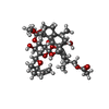
Mass: 680.823 Da / Num. of mol.: 1 / Source method: obtained synthetically / Formula: C36H56O12 / Feature type: SUBJECT OF INVESTIGATION
#4: Chemical ChemComp-MG / MAGNESIUM ION


Mass: 24.305 Da / Num. of mol.: 1 / Source method: obtained synthetically / Formula: Mg
#5: Water ChemComp-HOH / water / Water


Mass: 18.015 Da / Num. of mol.: 214 / Source method: isolated from a natural source / Formula: H2O
Has ligand of interestY

-
Experimental details

-
Experiment

ExperimentMethod: X-RAY DIFFRACTION / Number of used crystals: 1

-
Sample preparation

CrystalDensity Matthews: 2.67 Å3/Da / Density % sol: 53.89 %
Crystal growTemperature: 277 K / Method: vapor diffusion, hanging drop
Details: HEPES, magnesium chloride, calcium chloride, 2-mercaptoethanol, PEG400, glycerol, DMSO

-
Data collection

DiffractionMean temperature: 100 K / Serial crystal experiment: N
Diffraction sourceSource: SYNCHROTRON / Site: ESRF / Beamline: ID23-1 / Wavelength: 0.972425 Å
DetectorType: DECTRIS PILATUS 6M / Detector: PIXEL / Date: Dec 1, 2022
RadiationProtocol: SINGLE WAVELENGTH / Monochromatic (M) / Laue (L): M / Scattering type: x-ray
Radiation wavelengthWavelength: 0.972425 Å / Relative weight: 1
ReflectionResolution: 1.6→66.51 Å / Num. obs: 73894 / % possible obs: 100 % / Redundancy: 13.3 % / Biso Wilson estimate: 19.8 Å2 / CC1/2: 0.999 / Net I/σ(I): 30.1
Reflection shellResolution: 1.6→1.63 Å / Mean I/σ(I) obs: 7.1 / Num. unique obs: 1879 / CC1/2: 0.987

-
Processing

Software
NameVersionClassification
Aimlessdata scaling
MOLREPphasing
Cootmodel building
REFMAC5refinement
PHENIX1.20.1-4487refinement
RefinementMethod to determine structure: MOLECULAR REPLACEMENT / Resolution: 1.6→34.54 Å / SU ML: 0.1442 / Cross valid method: FREE R-VALUE / σ(F): 1.34 / Phase error: 20.3235
Stereochemistry target values: GeoStd + Monomer Library + CDL v1.2
RfactorNum. reflection% reflection
Rfree0.1996 3840 5.2 %
Rwork0.1916 70054 -
obs0.192 73894 99.98 %
Solvent computationShrinkage radii: 0.9 Å / VDW probe radii: 1.1 Å / Solvent model: FLAT BULK SOLVENT MODEL
Displacement parametersBiso mean: 24.06 Å2
Refinement stepCycle: LAST / Resolution: 1.6→34.54 Å
ProteinNucleic acidLigandSolventTotal
Num. atoms1831 0 49 214 2094
Refine LS restraints
Refine-IDTypeDev idealNumber
X-RAY DIFFRACTIONf_bond_d0.01242012
X-RAY DIFFRACTIONf_angle_d1.42522739
X-RAY DIFFRACTIONf_chiral_restr0.0854308
X-RAY DIFFRACTIONf_plane_restr0.0071355
X-RAY DIFFRACTIONf_dihedral_angle_d15.7635744
LS refinement shell
Resolution (Å)Rfactor RfreeNum. reflection RfreeRfactor RworkNum. reflection RworkRefine-ID% reflection obs (%)
1.6-1.620.19081430.23532615X-RAY DIFFRACTION100
1.62-1.640.231440.21262603X-RAY DIFFRACTION100
1.64-1.660.23311430.20622569X-RAY DIFFRACTION99.82
1.66-1.690.19911430.21772581X-RAY DIFFRACTION99.96
1.69-1.710.28121430.212579X-RAY DIFFRACTION100
1.71-1.740.24181420.2062620X-RAY DIFFRACTION100
1.74-1.770.20471400.20432613X-RAY DIFFRACTION100
1.77-1.80.22481420.20782573X-RAY DIFFRACTION99.93
1.8-1.830.20771450.19792620X-RAY DIFFRACTION100
1.83-1.870.2041390.20832568X-RAY DIFFRACTION100
1.87-1.90.20061390.20722598X-RAY DIFFRACTION100
1.9-1.950.22161400.21352601X-RAY DIFFRACTION100
1.95-1.990.22071460.21022587X-RAY DIFFRACTION100
1.99-2.040.20951420.20262590X-RAY DIFFRACTION100
2.04-2.10.19971380.18382573X-RAY DIFFRACTION100
2.1-2.160.19931450.18612628X-RAY DIFFRACTION100
2.16-2.230.20061430.18032602X-RAY DIFFRACTION100
2.23-2.310.16341400.18292588X-RAY DIFFRACTION100
2.31-2.40.20541440.18932584X-RAY DIFFRACTION100
2.4-2.510.23921400.19722604X-RAY DIFFRACTION100
2.51-2.640.19051440.18952555X-RAY DIFFRACTION100
2.64-2.810.22231440.19982610X-RAY DIFFRACTION100
2.81-3.020.20241460.19992620X-RAY DIFFRACTION100
3.02-3.330.18911400.18472595X-RAY DIFFRACTION100
3.33-3.810.20741390.18662576X-RAY DIFFRACTION100
3.81-4.790.16651440.16622595X-RAY DIFFRACTION100
4.8-34.540.18471420.19562607X-RAY DIFFRACTION99.93

+
About Yorodumi

-
News

-
Feb 9, 2022. New format data for meta-information of EMDB entries

New format data for meta-information of EMDB entries

  • Version 3 of the EMDB header file is now the official format.
  • The previous official version 1.9 will be removed from the archive.

Related info.:EMDB header

External links:wwPDB to switch to version 3 of the EMDB data model

-
Aug 12, 2020. Covid-19 info

Covid-19 info

URL: https://pdbj.org/emnavi/covid19.php

New page: Covid-19 featured information page in EM Navigator.

Related info.:Covid-19 info / Mar 5, 2020. Novel coronavirus structure data

+
Mar 5, 2020. Novel coronavirus structure data

Novel coronavirus structure data

Related info.:Yorodumi Speices / Aug 12, 2020. Covid-19 info

External links:COVID-19 featured content - PDBj / Molecule of the Month (242):Coronavirus Proteases

+
Jan 31, 2019. EMDB accession codes are about to change! (news from PDBe EMDB page)

EMDB accession codes are about to change! (news from PDBe EMDB page)

  • The allocation of 4 digits for EMDB accession codes will soon come to an end. Whilst these codes will remain in use, new EMDB accession codes will include an additional digit and will expand incrementally as the available range of codes is exhausted. The current 4-digit format prefixed with “EMD-” (i.e. EMD-XXXX) will advance to a 5-digit format (i.e. EMD-XXXXX), and so on. It is currently estimated that the 4-digit codes will be depleted around Spring 2019, at which point the 5-digit format will come into force.
  • The EM Navigator/Yorodumi systems omit the EMD- prefix.

Related info.:Q: What is EMD? / ID/Accession-code notation in Yorodumi/EM Navigator

External links:EMDB Accession Codes are Changing Soon! / Contact to PDBj

+
Jul 12, 2017. Major update of PDB

Major update of PDB

  • wwPDB released updated PDB data conforming to the new PDBx/mmCIF dictionary.
  • This is a major update changing the version number from 4 to 5, and with Remediation, in which all the entries are updated.
  • In this update, many items about electron microscopy experimental information are reorganized (e.g. em_software).
  • Now, EM Navigator and Yorodumi are based on the updated data.

External links:wwPDB Remediation / Enriched Model Files Conforming to OneDep Data Standards Now Available in the PDB FTP Archive

-
Yorodumi

Thousand views of thousand structures

  • Yorodumi is a browser for structure data from EMDB, PDB, SASBDB, etc.
  • This page is also the successor to EM Navigator detail page, and also detail information page/front-end page for Omokage search.
  • The word "yorodu" (or yorozu) is an old Japanese word meaning "ten thousand". "mi" (miru) is to see.

Related info.:EMDB / PDB / SASBDB / Comparison of 3 databanks / Yorodumi Search / Aug 31, 2016. New EM Navigator & Yorodumi / Yorodumi Papers / Jmol/JSmol / Function and homology information / Changes in new EM Navigator and Yorodumi

Read more