 Movie
Movie Controller

[English] 日本語
 Yorodumi
- PDB-8c27: Domain 3 of Group B Streptococcus pilus protein as scaffold for t... -
+ Open data

- Basic information
| Entry | Database: PDB / ID: 8c27 | ||||||
|---|---|---|---|---|---|---|---|
| Title | Domain 3 of Group B Streptococcus pilus protein as scaffold for the display of foreign epitopes | ||||||
 Components Components | PI-2a backbone protein | ||||||
 Keywords Keywords | MEMBRANE PROTEIN / Pilus protein / scaffold / epitopes. | ||||||
| Function / homology |  Function and homology information Function and homology information | ||||||
| Biological species |  Streptococcus agalactiae (bacteria) Streptococcus agalactiae (bacteria) | ||||||
| Method |  X-RAY DIFFRACTION / SYNCHROTRON / MOLECULAR REPLACEMENT / Resolution: 2.63 Å X-RAY DIFFRACTION / SYNCHROTRON / MOLECULAR REPLACEMENT / Resolution: 2.63 Å | ||||||
 Authors Authors | Veggi, D. / Cappelli, L. / Cozzi, R. | ||||||
| Funding support | 1items
| ||||||
 Citation Citation | Journal: Faseb J. / Year: 2024 Title: Computational structure-based approach to study chimeric antigens using a new protein scaffold displaying foreign epitopes. Authors: Cappelli, L. / Cinelli, P. / Perrotta, A. / Veggi, D. / Audagnotto, M. / Tuscano, G. / Pansegrau, W. / Bartolini, E. / Rinaudo, D. / Cozzi, R. | ||||||
| History |
|
- Structure visualization
| Structure viewer | Molecule: MolmilJmol/JSmol |
|---|
- Downloads & links
-Download
| PDBx/mmCIF format | 8c27.cif.gz | 68.8 KB | Display | PDBx/mmCIF format |
|---|---|---|---|---|
| PDB format | pdb8c27.ent.gz | 46.5 KB | Display | PDB format |
| PDBx/mmJSON format | 8c27.json.gz | Tree view | PDBx/mmJSON format | |
| Others |  Other downloads Other downloads |
-Validation report
| Arichive directory | https://data.pdbj.org/pub/pdb/validation_reports/c2/8c27ftp://data.pdbj.org/pub/pdb/validation_reports/c2/8c27 | HTTPS FTP |
|---|
-Related structure data
| Related structure data | 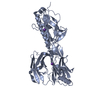 2xtlS S: Starting model for refinement |
|---|---|
| Similar structure data |
-Links
 PDBj
PDBj- Assembly
| Deposited unit | 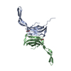
| ||||||||||||
|---|---|---|---|---|---|---|---|---|---|---|---|---|---|
| 1 | 
| ||||||||||||
| 2 | 
| ||||||||||||
| Unit cell |
|
-Components
| #1: Protein | Mass: 14666.284 Da / Num. of mol.: 2 Source method: isolated from a genetically manipulated source Source: (gene. exp.) Streptococcus agalactiae (bacteria)Production host: References: UniProt: B9UR22 #2: Chemical |   Mass: 62.068 Da / Num. of mol.: 2 / Source method: obtained synthetically / Formula: C2H6O2 / Feature type: SUBJECT OF INVESTIGATION Mass: 62.068 Da / Num. of mol.: 2 / Source method: obtained synthetically / Formula: C2H6O2 / Feature type: SUBJECT OF INVESTIGATION#3: Water | ChemComp-HOH / |  Mass: 18.015 Da / Num. of mol.: 6 / Source method: isolated from a natural source / Formula: H2O Mass: 18.015 Da / Num. of mol.: 6 / Source method: isolated from a natural source / Formula: H2OHas ligand of interest | Y | |
|---|
-Experimental details
-Experiment
| Experiment | Method: X-RAY DIFFRACTION / Number of used crystals: 1 |
|---|
- Sample preparation
| Crystal | Density Matthews: 3.41 Å3/Da / Density % sol: 63.91 % |
|---|---|
| Crystal grow | Temperature: 291 K / Method: vapor diffusion, sitting drop / Details: 0.1M HEPES with 20% w/v jeff ED-2001 pH 6.5 |
-Data collection
| Diffraction | Mean temperature: 100 K / Serial crystal experiment: N |
|---|---|
| Diffraction source | Source: SYNCHROTRON / Site: ESRF  / Beamline: ID30B / Wavelength: 0.96546 Å / Beamline: ID30B / Wavelength: 0.96546 Å |
| Detector | Type: DECTRIS PILATUS 2M / Detector: PIXEL / Date: Sep 17, 2021 |
| Radiation | Protocol: SINGLE WAVELENGTH / Monochromatic (M) / Laue (L): M / Scattering type: x-ray |
| Radiation wavelength | Wavelength: 0.96546 Å / Relative weight: 1 |
| Reflection | Resolution: 2.63→94.9 Å / Num. obs: 12062 / % possible obs: 99.7 % / Redundancy: 5.8 % / Biso Wilson estimate: 60.74 Å2 / CC1/2: 0.998 / Net I/σ(I): 13.5 |
| Reflection shell | Resolution: 2.63→94.9 Å / Num. unique obs: 12062 / CC1/2: 0.998 |
- Processing
| Software |
| |||||||||||||||||||||||||||||||||||
|---|---|---|---|---|---|---|---|---|---|---|---|---|---|---|---|---|---|---|---|---|---|---|---|---|---|---|---|---|---|---|---|---|---|---|---|---|
| Refinement | Method to determine structure: MOLECULAR REPLACEMENT Starting model: 2XTL Resolution: 2.63→58.15 Å / SU ML: 0.4785 / Cross valid method: FREE R-VALUE / σ(F): 1.35 / Phase error: 28.1437 Stereochemistry target values: GeoStd + Monomer Library + CDL v1.2
| |||||||||||||||||||||||||||||||||||
| Solvent computation | Shrinkage radii: 0.9 Å / VDW probe radii: 1.1 Å / Solvent model: FLAT BULK SOLVENT MODEL | |||||||||||||||||||||||||||||||||||
| Displacement parameters | Biso mean: 56.1 Å2 | |||||||||||||||||||||||||||||||||||
| Refinement step | Cycle: LAST / Resolution: 2.63→58.15 Å
| |||||||||||||||||||||||||||||||||||
| Refine LS restraints |
| |||||||||||||||||||||||||||||||||||
| LS refinement shell |
|
