 Movie
Movie Controller


+ Open data

- Basic information
| Entry | Database: PDB / ID: 8byr | ||||||
|---|---|---|---|---|---|---|---|
| Title | Crystal structure of MoaB2 protein from Mycobacterium smegmatis | ||||||
 Components Components | Molybdopterin biosynthesis protein | ||||||
 Keywords Keywords | BIOSYNTHETIC PROTEIN / Sigma factor binding protein | ||||||
| Function / homology | : / MoaB/Mog domain / MoaB/Mog-like domain superfamily / Probable molybdopterin binding domain / Probable molybdopterin binding domain / Mo-molybdopterin cofactor biosynthetic process / Molybdopterin biosynthesis protein Function and homology information Function and homology information | ||||||
| Biological species |  Mycolicibacterium smegmatis (bacteria) Mycolicibacterium smegmatis (bacteria) | ||||||
| Method |  X-RAY DIFFRACTION / SYNCHROTRON / MOLECULAR REPLACEMENT / Resolution: 2.53 Å X-RAY DIFFRACTION / SYNCHROTRON / MOLECULAR REPLACEMENT / Resolution: 2.53 Å | ||||||
 Authors Authors | Narasimhan, S. / Sanderova, H. / Zidek, L. / Krasny, L. | ||||||
| Funding support |  Czech Republic, 1items Czech Republic, 1items
| ||||||
 Citation Citation | Journal: J.Bacteriol. / Year: 2024 Title: MoaB2, a newly identified transcription factor, binds to sigma A in Mycobacterium smegmatis. Authors: Brezovska, B. / Narasimhan, S. / Sikova, M. / Sanderova, H. / Koval, T. / Borah, N. / Shoman, M. / Pospisilova, D. / Vankova Hausnerova, V. / Tuzincin, D. / Cerny, M. / Komarek, J. / ...Authors: Brezovska, B. / Narasimhan, S. / Sikova, M. / Sanderova, H. / Koval, T. / Borah, N. / Shoman, M. / Pospisilova, D. / Vankova Hausnerova, V. / Tuzincin, D. / Cerny, M. / Komarek, J. / Janouskova, M. / Kambova, M. / Halada, P. / Krenkova, A. / Hubalek, M. / Trundova, M. / Dohnalek, J. / Hnilicova, J. / Zidek, L. / Krasny, L. | ||||||
| History |
|
- Structure visualization
| Structure viewer | Molecule: MolmilJmol/JSmol |
|---|
- Downloads & links
-Download
| PDBx/mmCIF format | 8byr.cif.gz | 166.3 KB | Display | PDBx/mmCIF format |
|---|---|---|---|---|
| PDB format | pdb8byr.ent.gz | 131.6 KB | Display | PDB format |
| PDBx/mmJSON format | 8byr.json.gz | Tree view | PDBx/mmJSON format | |
| Others |  Other downloads Other downloads |
-Validation report
| Arichive directory | https://data.pdbj.org/pub/pdb/validation_reports/by/8byrftp://data.pdbj.org/pub/pdb/validation_reports/by/8byr | HTTPS FTP |
|---|
-Related structure data
| Related structure data | 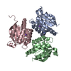 3rfqS S: Starting model for refinement |
|---|---|
| Similar structure data |
-Links
 PDBj
PDBj- Assembly
| Deposited unit | 
| ||||||||
|---|---|---|---|---|---|---|---|---|---|
| 1 |
| ||||||||
| Unit cell |
|
-Components
| #1: Protein | Mass: 17621.857 Da / Num. of mol.: 6 Source method: isolated from a genetically manipulated source Source: (gene. exp.) Mycolicibacterium smegmatis (bacteria)Strain: Mycolicibacterium smegmatis (strain ATCC 700084 / mc(2)155) (Mycobacterium smegmatis) Gene: MSMEG_5485 / Plasmid: pET22b(+) / Production host: #2: Water | ChemComp-HOH / |  Mass: 18.015 Da / Num. of mol.: 101 / Source method: isolated from a natural source / Formula: H2O Mass: 18.015 Da / Num. of mol.: 101 / Source method: isolated from a natural source / Formula: H2OHas protein modification | N | |
|---|
-Experimental details
-Experiment
| Experiment | Method: X-RAY DIFFRACTION / Number of used crystals: 1 |
|---|
- Sample preparation
| Crystal | Density Matthews: 2.45 Å3/Da / Density % sol: 49.93 % |
|---|---|
| Crystal grow | Temperature: 293.15 K / Method: vapor diffusion, sitting drop / pH: 7.5 Details: 0.2M calcium chloride, 14% w/v PEG 400, 0.1M sodium-HEPES (pH 7.5) |
-Data collection
| Diffraction | Mean temperature: 100 K / Serial crystal experiment: N |
|---|---|
| Diffraction source | Source: SYNCHROTRON / Site: PETRA III, EMBL c/o DESY  / Beamline: P14 (MX2) / Wavelength: 0.9762 Å / Beamline: P14 (MX2) / Wavelength: 0.9762 Å |
| Detector | Type: DECTRIS EIGER2 X CdTe 16M / Detector: PIXEL / Date: Nov 9, 2020 / Details: Toroidal mirror |
| Radiation | Monochromator: Si(111) / Protocol: SINGLE WAVELENGTH / Monochromatic (M) / Laue (L): M / Scattering type: x-ray |
| Radiation wavelength | Wavelength: 0.9762 Å / Relative weight: 1 |
| Reflection | Resolution: 2.53→47.69 Å / Num. obs: 35068 / % possible obs: 99.9 % / Redundancy: 13.4 % / CC1/2: 0.999 / Rmerge(I) obs: 0.136 / Rpim(I) all: 0.038 / Rrim(I) all: 0.141 / Χ2: 0.99 / Net I/σ(I): 15 / Num. measured all: 470613 |
| Reflection shell | Resolution: 2.53→2.64 Å / % possible obs: 99.5 % / Redundancy: 13.4 % / Rmerge(I) obs: 0.868 / Num. measured all: 56564 / Num. unique obs: 4206 / CC1/2: 0.872 / Rpim(I) all: 0.242 / Rrim(I) all: 0.902 / Χ2: 0.98 / Net I/σ(I) obs: 3.4 |
- Processing
| Software |
| ||||||||||||||||||||||||||||||||||||||||||||||||||||||||||||||||||||||||||||||||||||||||||||||||||||||||||||||||||||||||||||||||||||||||||||||||||||||||||||||||||||||||||||||||||||||
|---|---|---|---|---|---|---|---|---|---|---|---|---|---|---|---|---|---|---|---|---|---|---|---|---|---|---|---|---|---|---|---|---|---|---|---|---|---|---|---|---|---|---|---|---|---|---|---|---|---|---|---|---|---|---|---|---|---|---|---|---|---|---|---|---|---|---|---|---|---|---|---|---|---|---|---|---|---|---|---|---|---|---|---|---|---|---|---|---|---|---|---|---|---|---|---|---|---|---|---|---|---|---|---|---|---|---|---|---|---|---|---|---|---|---|---|---|---|---|---|---|---|---|---|---|---|---|---|---|---|---|---|---|---|---|---|---|---|---|---|---|---|---|---|---|---|---|---|---|---|---|---|---|---|---|---|---|---|---|---|---|---|---|---|---|---|---|---|---|---|---|---|---|---|---|---|---|---|---|---|---|---|---|---|
| Refinement | Method to determine structure: MOLECULAR REPLACEMENT Starting model: 3RFQ Resolution: 2.53→47.69 Å / Cor.coef. Fo:Fc: 0.951 / Cor.coef. Fo:Fc free: 0.911 / SU B: 11.207 / SU ML: 0.233 / Cross valid method: THROUGHOUT / ESU R: 0.45 / ESU R Free: 0.294 / Stereochemistry target values: MAXIMUM LIKELIHOOD / Details: HYDROGENS HAVE BEEN ADDED IN THE RIDING POSITIONS
| ||||||||||||||||||||||||||||||||||||||||||||||||||||||||||||||||||||||||||||||||||||||||||||||||||||||||||||||||||||||||||||||||||||||||||||||||||||||||||||||||||||||||||||||||||||||
| Solvent computation | Ion probe radii: 0.8 Å / Shrinkage radii: 0.8 Å / VDW probe radii: 1.2 Å / Solvent model: MASK | ||||||||||||||||||||||||||||||||||||||||||||||||||||||||||||||||||||||||||||||||||||||||||||||||||||||||||||||||||||||||||||||||||||||||||||||||||||||||||||||||||||||||||||||||||||||
| Displacement parameters | Biso mean: 49.976 Å2
| ||||||||||||||||||||||||||||||||||||||||||||||||||||||||||||||||||||||||||||||||||||||||||||||||||||||||||||||||||||||||||||||||||||||||||||||||||||||||||||||||||||||||||||||||||||||
| Refinement step | Cycle: 1 / Resolution: 2.53→47.69 Å
| ||||||||||||||||||||||||||||||||||||||||||||||||||||||||||||||||||||||||||||||||||||||||||||||||||||||||||||||||||||||||||||||||||||||||||||||||||||||||||||||||||||||||||||||||||||||
| Refine LS restraints |
|