 Movie
Movie Controller

[English] 日本語
 Yorodumi
- PDB-8by4: Crystal structure of Odorant Binding Protein 1 from Aedes albopic... -
+ Open data

- Basic information
| Entry | Database: PDB / ID: 8by4 | |||||||||
|---|---|---|---|---|---|---|---|---|---|---|
| Title | Crystal structure of Odorant Binding Protein 1 from Aedes albopictus (Asian tiger mosquito) | |||||||||
 Components Components | Odorant-binding protein 1 | |||||||||
 Keywords Keywords | TRANSPORT PROTEIN / Odorant Binding Protein (OBP) / mosquito / olfaction / Carvacrol | |||||||||
| Function / homology | : / Insect pheromone/odorant binding protein domains. / Pheromone/general odorant binding protein / PBP/GOBP family / Pheromone/general odorant binding protein superfamily / odorant binding / extracellular region / Odorant-binding protein 1 Function and homology information Function and homology information | |||||||||
| Biological species |  | |||||||||
| Method |  X-RAY DIFFRACTION / SYNCHROTRON / MOLECULAR REPLACEMENT / Resolution: 2 Å X-RAY DIFFRACTION / SYNCHROTRON / MOLECULAR REPLACEMENT / Resolution: 2 Å | |||||||||
 Authors Authors | Liggri, P.G.V. / Tsitsanou, K.E. / Zographos, S.E. | |||||||||
| Funding support |  Greece, 2items Greece, 2items
| |||||||||
 Citation Citation | Journal: To Be Published Title: Crystal structure of Odorant Binding Protein 1 from Aedes albopictus (Asian tiger mosquito) Authors: Liggri, P.G.V. / Tsitsanou, K.E. / Zographos, S.E. | |||||||||
| History |
|
- Structure visualization
| Structure viewer | Molecule: MolmilJmol/JSmol |
|---|
- Downloads & links
-Download
| PDBx/mmCIF format | 8by4.cif.gz | 66.6 KB | Display | PDBx/mmCIF format |
|---|---|---|---|---|
| PDB format | pdb8by4.ent.gz | 47.8 KB | Display | PDB format |
| PDBx/mmJSON format | 8by4.json.gz | Tree view | PDBx/mmJSON format | |
| Others |  Other downloads Other downloads |
-Validation report
| Summary document | 8by4_validation.pdf.gz | 409.2 KB | Display | wwPDB validaton report |
|---|---|---|---|---|
| Full document | 8by4_full_validation.pdf.gz | 409.2 KB | Display | |
| Data in XML | 8by4_validation.xml.gz | 7.1 KB | Display | |
| Data in CIF | 8by4_validation.cif.gz | 9 KB | Display | |
| Arichive directory | https://data.pdbj.org/pub/pdb/validation_reports/by/8by4ftp://data.pdbj.org/pub/pdb/validation_reports/by/8by4 | HTTPS FTP |
-Related structure data
| Related structure data | 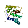 6og0S S: Starting model for refinement |
|---|---|
| Similar structure data |
-Links
 PDBj
PDBj- Assembly
| Deposited unit | 
| ||||||||
|---|---|---|---|---|---|---|---|---|---|
| 1 |
| ||||||||
| Unit cell |
| ||||||||
| Components on special symmetry positions |
|
-Components
| #1: Protein | Mass: 14190.069 Da / Num. of mol.: 1 Source method: isolated from a genetically manipulated source Source: (gene. exp.) Gene: 109422789, RP20_CCG014341, RP20_CCG027709 / Production host:  |
|---|---|
| #2: Water | ChemComp-HOH /  Mass: 18.015 Da / Num. of mol.: 58 / Source method: isolated from a natural source / Formula: H2O Mass: 18.015 Da / Num. of mol.: 58 / Source method: isolated from a natural source / Formula: H2O |
| Has protein modification | Y |
-Experimental details
-Experiment
| Experiment | Method: X-RAY DIFFRACTION / Number of used crystals: 1 |
|---|
- Sample preparation
| Crystal | Density Matthews: 4.3 Å3/Da / Density % sol: 71.37 % |
|---|---|
| Crystal grow | Temperature: 293 K / Method: vapor diffusion, sitting drop / pH: 6.5 / Details: 0.1.6M tri-sodium citrate |
-Data collection
| Diffraction | Mean temperature: 100 K / Serial crystal experiment: N |
|---|---|
| Diffraction source | Source: SYNCHROTRON / Site: Diamond  / Beamline: I04-1 / Wavelength: 0.9159 Å / Beamline: I04-1 / Wavelength: 0.9159 Å |
| Detector | Type: DECTRIS EIGER2 XE 9M / Detector: PIXEL / Date: Apr 6, 2019 |
| Radiation | Protocol: SINGLE WAVELENGTH / Monochromatic (M) / Laue (L): M / Scattering type: x-ray |
| Radiation wavelength | Wavelength: 0.9159 Å / Relative weight: 1 |
| Reflection | Resolution: 2→47.35 Å / Num. obs: 16836 / % possible obs: 99.96 % / Redundancy: 2 % / CC1/2: 0.999 / Net I/σ(I): 15.6 |
| Reflection shell | Resolution: 2→2.072 Å / Redundancy: 2 % / Mean I/σ(I) obs: 3.65 / Num. unique obs: 1662 / CC1/2: 0.94 / % possible all: 99.82 |
- Processing
| Software |
| |||||||||||||||||||||||||||||||||||||||||||||||||||||||||||||||||||||||||||||||||||||||||||||||||||||||||
|---|---|---|---|---|---|---|---|---|---|---|---|---|---|---|---|---|---|---|---|---|---|---|---|---|---|---|---|---|---|---|---|---|---|---|---|---|---|---|---|---|---|---|---|---|---|---|---|---|---|---|---|---|---|---|---|---|---|---|---|---|---|---|---|---|---|---|---|---|---|---|---|---|---|---|---|---|---|---|---|---|---|---|---|---|---|---|---|---|---|---|---|---|---|---|---|---|---|---|---|---|---|---|---|---|---|---|
| Refinement | Method to determine structure: MOLECULAR REPLACEMENT Starting model: 6OG0 Resolution: 2→47.35 Å / Cor.coef. Fo:Fc: 0.966 / Cor.coef. Fo:Fc free: 0.948 / SU B: 4.561 / SU ML: 0.064 / Cross valid method: THROUGHOUT / ESU R: 0.101 / ESU R Free: 0.104 / Stereochemistry target values: MAXIMUM LIKELIHOOD Details: U VALUES : WITH TLS ADDED HYDROGENS HAVE BEEN ADDED IN THE RIDING POSITIONS U VALUES : RESIDUAL ONLY
| |||||||||||||||||||||||||||||||||||||||||||||||||||||||||||||||||||||||||||||||||||||||||||||||||||||||||
| Solvent computation | Ion probe radii: 0.7 Å / Shrinkage radii: 0.7 Å / VDW probe radii: 1.2 Å / Solvent model: MASK | |||||||||||||||||||||||||||||||||||||||||||||||||||||||||||||||||||||||||||||||||||||||||||||||||||||||||
| Displacement parameters | Biso mean: 40.683 Å2
| |||||||||||||||||||||||||||||||||||||||||||||||||||||||||||||||||||||||||||||||||||||||||||||||||||||||||
| Refinement step | Cycle: LAST / Resolution: 2→47.35 Å
| |||||||||||||||||||||||||||||||||||||||||||||||||||||||||||||||||||||||||||||||||||||||||||||||||||||||||
| Refine LS restraints |
| |||||||||||||||||||||||||||||||||||||||||||||||||||||||||||||||||||||||||||||||||||||||||||||||||||||||||
| LS refinement shell | Resolution: 2→2.052 Å / Total num. of bins used: 20
| |||||||||||||||||||||||||||||||||||||||||||||||||||||||||||||||||||||||||||||||||||||||||||||||||||||||||
| Refinement TLS params. | Method: refined / Origin x: 26.774 Å / Origin y: -26.389 Å / Origin z: 5.905 Å
|
