[English] 日本語
 Yorodumi
- PDB-8bx8: In situ outer dynein arm from Chlamydomonas reinhardtii in the po... -
+ Open data
- Basic information
| Entry | Database: PDB / ID: 8bx8 | |||||||||||||||||||||||||||||||||
|---|---|---|---|---|---|---|---|---|---|---|---|---|---|---|---|---|---|---|---|---|---|---|---|---|---|---|---|---|---|---|---|---|---|---|
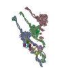
| Title | In situ outer dynein arm from Chlamydomonas reinhardtii in the post-power stroke state | |||||||||||||||||||||||||||||||||
 Components Components |
| |||||||||||||||||||||||||||||||||
 Keywords Keywords | MOTOR PROTEIN / axoneme / outer dynein arm / post power stroke / dynein | |||||||||||||||||||||||||||||||||
| Function / homology |  Function and homology information Function and homology informationaxonemal dynein complex / outer dynein arm / outer dynein arm assembly / dynein light chain binding / cilium movement / dynein heavy chain binding / dynein complex / myosin II complex / minus-end-directed microtubule motor activity / cytoplasmic dynein complex ...axonemal dynein complex / outer dynein arm / outer dynein arm assembly / dynein light chain binding / cilium movement / dynein heavy chain binding / dynein complex / myosin II complex / minus-end-directed microtubule motor activity / cytoplasmic dynein complex / dynein light intermediate chain binding / motile cilium / microtubule-based movement / dynein intermediate chain binding / microtubule-based process / cell projection / microtubule / cilium / calcium ion binding / ATP hydrolysis activity / ATP binding / cytoplasm Similarity search - Function | |||||||||||||||||||||||||||||||||
| Biological species |  | |||||||||||||||||||||||||||||||||
| Method | ELECTRON MICROSCOPY / subtomogram averaging / cryo EM / Resolution: 30.3 Å | |||||||||||||||||||||||||||||||||
 Authors Authors | Zimmermann, N.E.L. / Noga, A. / Obbineni, J.M. / Ishikawa, T. | |||||||||||||||||||||||||||||||||
| Funding support |  Switzerland, 1items Switzerland, 1items
| |||||||||||||||||||||||||||||||||
 Citation Citation |  Journal: EMBO J / Year: 2023 Journal: EMBO J / Year: 2023Title: ATP-induced conformational change of axonemal outer dynein arms revealed by cryo-electron tomography. Authors: Noemi Zimmermann / Akira Noga / Jagan Mohan Obbineni / Takashi Ishikawa /   Abstract: Axonemal outer dynein arm (ODA) motors generate force for ciliary beating. We analyzed three states of the ODA during the power stroke cycle using in situ cryo-electron tomography, subtomogram ...Axonemal outer dynein arm (ODA) motors generate force for ciliary beating. We analyzed three states of the ODA during the power stroke cycle using in situ cryo-electron tomography, subtomogram averaging, and classification. These states of force generation depict the prepower stroke, postpower stroke, and intermediate state conformations. Comparison of these conformations to published in vitro atomic structures of cytoplasmic dynein, ODA, and the Shulin-ODA complex revealed differences in the orientation and position of the dynein head. Our analysis shows that in the absence of ATP, all dynein linkers interact with the AAA3/AAA4 domains, indicating that interactions with the adjacent microtubule doublet B-tubule direct dynein orientation. For the prepower stroke conformation, there were changes in the tail that is anchored on the A-tubule. We built models starting with available high-resolution structures to generate a best-fitting model structure for the in situ pre- and postpower stroke ODA conformations, thereby showing that ODA in a complex with Shulin adopts a similar conformation as the active prepower stroke ODA in the axoneme. | |||||||||||||||||||||||||||||||||
| History |
|
- Structure visualization
| Structure viewer | Molecule:  Molmil Molmil Jmol/JSmol Jmol/JSmol |
|---|
- Downloads & links
- Download
| PDBx/mmCIF format |  8bx8.cif.gz 8bx8.cif.gz | 2.9 MB | Display |  PDBx/mmCIF format PDBx/mmCIF format |
|---|---|---|---|---|
| PDB format |  pdb8bx8.ent.gz pdb8bx8.ent.gz | Display |  PDB format PDB format | |
| PDBx/mmJSON format |  8bx8.json.gz 8bx8.json.gz | Tree view |  PDBx/mmJSON format PDBx/mmJSON format | |
| Others |  Other downloads Other downloads |
-Validation report
| Summary document |  8bx8_validation.pdf.gz 8bx8_validation.pdf.gz | 1.9 MB | Display |  wwPDB validaton report wwPDB validaton report |
|---|---|---|---|---|
| Full document |  8bx8_full_validation.pdf.gz 8bx8_full_validation.pdf.gz | 2.8 MB | Display | |
| Data in XML |  8bx8_validation.xml.gz 8bx8_validation.xml.gz | 471 KB | Display | |
| Data in CIF |  8bx8_validation.cif.gz 8bx8_validation.cif.gz | 721.2 KB | Display | |
| Arichive directory |  https://data.pdbj.org/pub/pdb/validation_reports/bx/8bx8 https://data.pdbj.org/pub/pdb/validation_reports/bx/8bx8 ftp://data.pdbj.org/pub/pdb/validation_reports/bx/8bx8 ftp://data.pdbj.org/pub/pdb/validation_reports/bx/8bx8 | HTTPS FTP |
-Related structure data
| Related structure data |  16312MC  8bwyC M: map data used to model this data C: citing same article ( |
|---|---|
| Similar structure data | Similarity search - Function & homology  F&H Search F&H Search |
- Links
Links
- Assembly
| Deposited unit | 
|
|---|---|
| 1 |
|
- Components
-Protein , 5 types, 5 molecules ACDEP
| #1: Protein | Mass: 534400.938 Da / Num. of mol.: 1 / Source method: isolated from a natural source / Source: (natural)  |
|---|---|
| #3: Protein | Mass: 475554.406 Da / Num. of mol.: 1 / Source method: isolated from a natural source / Source: (natural)  |
| #4: Protein | Mass: 76685.891 Da / Num. of mol.: 1 / Source method: isolated from a natural source / Source: (natural)  |
| #5: Protein | Mass: 77178.062 Da / Num. of mol.: 1 / Source method: isolated from a natural source / Source: (natural)  |
| #16: Protein | Mass: 14256.472 Da / Num. of mol.: 1 / Source method: isolated from a natural source / Source: (natural)  |
-Dynein light ... , 12 types, 12 molecules FGHIJKLMNOQR
| #6: Protein | Mass: 14751.817 Da / Num. of mol.: 1 / Source method: isolated from a natural source / Source: (natural)  |
|---|---|
| #7: Protein | Mass: 18490.188 Da / Num. of mol.: 1 / Source method: isolated from a natural source / Source: (natural)  |
| #8: Protein | Mass: 10780.357 Da / Num. of mol.: 1 / Source method: isolated from a natural source / Source: (natural)  |
| #9: Protein | Mass: 12348.086 Da / Num. of mol.: 1 / Source method: isolated from a natural source / Source: (natural)  |
| #10: Protein | Mass: 11435.072 Da / Num. of mol.: 1 / Source method: isolated from a natural source / Source: (natural)  |
| #11: Protein | Mass: 10973.408 Da / Num. of mol.: 1 / Source method: isolated from a natural source / Source: (natural)  |
| #12: Protein | Mass: 12516.457 Da / Num. of mol.: 1 / Source method: isolated from a natural source / Source: (natural)  |
| #13: Protein | Mass: 10453.167 Da / Num. of mol.: 1 / Source method: isolated from a natural source / Source: (natural)  |
| #14: Protein | Mass: 13202.817 Da / Num. of mol.: 1 / Source method: isolated from a natural source / Source: (natural)  |
| #15: Protein | Mass: 15608.120 Da / Num. of mol.: 1 / Source method: isolated from a natural source / Source: (natural)  |
| #17: Protein | Mass: 22851.654 Da / Num. of mol.: 1 / Source method: isolated from a natural source / Source: (natural)  |
| #18: Protein | Mass: 17822.795 Da / Num. of mol.: 1 / Source method: isolated from a natural source / Source: (natural)  |
-Antibody , 1 types, 1 molecules B
| #2: Antibody | Mass: 530182.375 Da / Num. of mol.: 1 / Source method: isolated from a natural source / Source: (natural)  |
|---|
-Non-polymers , 3 types, 18 molecules 


| #19: Chemical | ChemComp-ADP / #20: Chemical | #21: Chemical | ChemComp-MG / |
|---|
-Details
| Has ligand of interest | N |
|---|---|
| Has protein modification | Y |
-Experimental details
-Experiment
| Experiment | Method: ELECTRON MICROSCOPY |
|---|---|
| EM experiment | Aggregation state: PARTICLE / 3D reconstruction method: subtomogram averaging |
- Sample preparation
| Component | Name: In situ outer dynein arm / Type: ORGANELLE OR CELLULAR COMPONENT / Entity ID: #1-#18 / Source: NATURAL |
|---|---|
| Molecular weight | Experimental value: NO |
| Source (natural) | Organism:  |
| Buffer solution | pH: 7.4 |
| Specimen | Embedding applied: NO / Shadowing applied: NO / Staining applied: NO / Vitrification applied: YES |
| Vitrification | Cryogen name: ETHANE |
- Electron microscopy imaging
| Experimental equipment |  Model: Titan Krios / Image courtesy: FEI Company |
|---|---|
| Microscopy | Model: FEI TITAN KRIOS |
| Electron gun | Electron source:  FIELD EMISSION GUN / Accelerating voltage: 300 kV / Illumination mode: FLOOD BEAM FIELD EMISSION GUN / Accelerating voltage: 300 kV / Illumination mode: FLOOD BEAM |
| Electron lens | Mode: BRIGHT FIELD / Nominal defocus max: 4000 nm / Nominal defocus min: 3000 nm |
| Image recording | Electron dose: 1 e/Å2 / Avg electron dose per subtomogram: 80 e/Å2 / Film or detector model: GATAN K2 QUANTUM (4k x 4k) |
- Processing
| Software | Name: UCSF ChimeraX / Version: 1.4/v9 / Classification: model building / URL: https://www.rbvi.ucsf.edu/chimerax/ / Os: Windows / Type: package |
|---|---|
| CTF correction | Type: PHASE FLIPPING ONLY |
| Symmetry | Point symmetry: C1 (asymmetric) |
| 3D reconstruction | Resolution: 30.3 Å / Resolution method: FSC 0.143 CUT-OFF / Num. of particles: 2131 / Symmetry type: POINT |
| EM volume selection | Num. of tomograms: 8 / Num. of volumes extracted: 3167 |
| Atomic model building | Protocol: RIGID BODY FIT / Space: REAL |
 Movie
Movie Controller


 PDBj
PDBj


