 Movie
Movie Controller


+ Open data

- Basic information
| Entry | Database: PDB / ID: 8bx1 | ||||||
|---|---|---|---|---|---|---|---|
| Title | Oct4/Sox2 protein:DNA complex | ||||||
 Components Components |
| ||||||
 Keywords Keywords | TRANSCRIPTION / Oct4 / Sox2 / Hoxb1 | ||||||
| Function / homology |  Function and homology information Function and homology informationdiencephalon morphogenesis / olfactory placode formation / lens induction in camera-type eye / ectodermal cell fate commitment / response to benzoic acid / retina morphogenesis in camera-type eye / mesodermal cell fate commitment / HMG box domain binding / detection of mechanical stimulus involved in equilibrioception / pigment biosynthetic process ...diencephalon morphogenesis / olfactory placode formation / lens induction in camera-type eye / ectodermal cell fate commitment / response to benzoic acid / retina morphogenesis in camera-type eye / mesodermal cell fate commitment / HMG box domain binding / detection of mechanical stimulus involved in equilibrioception / pigment biosynthetic process / trophectodermal cell fate commitment / forebrain neuron differentiation / anatomical structure formation involved in morphogenesis / regulation of asymmetric cell division / endodermal cell fate specification / positive regulation of epithelial cell differentiation / adenohypophysis development / tongue development / Deactivation of the beta-catenin transactivating complex / blastocyst growth / POU domain binding / endodermal cell fate commitment / germ-line stem cell population maintenance / neuron fate commitment / neuronal stem cell population maintenance / negative regulation of calcium ion-dependent exocytosis / trophectodermal cell differentiation / detection of mechanical stimulus involved in sensory perception of sound / male genitalia development / female germ cell nucleus / cell fate specification / germ cell nucleus / RNA polymerase II intronic transcription regulatory region sequence-specific DNA binding / inner ear morphogenesis / lung alveolus development / stem cell population maintenance / positive regulation of neuroblast proliferation / negative regulation of neuron differentiation / positive regulation of Notch signaling pathway / epithelial tube branching involved in lung morphogenesis / cytokine binding / cellular response to cadmium ion / negative regulation of Wnt signaling pathway / blastocyst development / somatic stem cell population maintenance / negative regulation of cell differentiation / neuroblast proliferation / cell fate commitment / embryonic organ development / regulation of neurogenesis / positive regulation of transcription initiation by RNA polymerase II / response to retinoic acid / negative regulation of osteoblast differentiation / cis-regulatory region sequence-specific DNA binding / : / Notch signaling pathway / transcription repressor complex / positive regulation of neuron differentiation / negative regulation of phosphatidylinositol 3-kinase/protein kinase B signal transduction / telomere maintenance / cellular response to leukemia inhibitory factor / male germ cell nucleus / stem cell differentiation / sensory perception of sound / negative regulation of canonical Wnt signaling pathway / cerebral cortex development / chromatin DNA binding / Wnt signaling pathway / nuclear receptor activity / neuron differentiation / osteoblast differentiation / DNA-binding transcription activator activity, RNA polymerase II-specific / transcription regulator complex / gene expression / sequence-specific DNA binding / DNA-binding transcription factor binding / RNA polymerase II-specific DNA-binding transcription factor binding / transcription by RNA polymerase II / DNA-binding transcription factor activity, RNA polymerase II-specific / positive regulation of phosphatidylinositol 3-kinase/protein kinase B signal transduction / transcription cis-regulatory region binding / RNA polymerase II cis-regulatory region sequence-specific DNA binding / DNA-binding transcription factor activity / negative regulation of gene expression / negative regulation of DNA-templated transcription / ubiquitin protein ligase binding / chromatin binding / positive regulation of gene expression / regulation of DNA-templated transcription / regulation of transcription by RNA polymerase II / chromatin / positive regulation of DNA-templated transcription / nucleolus / negative regulation of transcription by RNA polymerase II / positive regulation of transcription by RNA polymerase II / DNA binding / nucleoplasm / nucleus / cytoplasm Similarity search - Function | ||||||
| Biological species |  | ||||||
| Method |  X-RAY DIFFRACTION / SYNCHROTRON / MOLECULAR REPLACEMENT / Resolution: 2.5 Å X-RAY DIFFRACTION / SYNCHROTRON / MOLECULAR REPLACEMENT / Resolution: 2.5 Å | ||||||
 Authors Authors | Chojnowski, G. / Wilmanns, M. / Round, E. | ||||||
| Funding support | 1items
| ||||||
 Citation Citation | Journal: To Be Published Title: Crystal structure of Oct4-Soc2-HoxB1 complex Authors: Chojnowski, G. / Wilmanns, M. / Round, E. | ||||||
| History |
|
- Structure visualization
| Structure viewer | Molecule: MolmilJmol/JSmol |
|---|
- Downloads & links
-Download
| PDBx/mmCIF format | 8bx1.cif.gz | 147.6 KB | Display | PDBx/mmCIF format |
|---|---|---|---|---|
| PDB format | pdb8bx1.ent.gz | 110.7 KB | Display | PDB format |
| PDBx/mmJSON format | 8bx1.json.gz | Tree view | PDBx/mmJSON format | |
| Others |  Other downloads Other downloads |
-Validation report
| Summary document | 8bx1_validation.pdf.gz | 447.9 KB | Display | wwPDB validaton report |
|---|---|---|---|---|
| Full document | 8bx1_full_validation.pdf.gz | 452.5 KB | Display | |
| Data in XML | 8bx1_validation.xml.gz | 11 KB | Display | |
| Data in CIF | 8bx1_validation.cif.gz | 14.2 KB | Display | |
| Arichive directory | https://data.pdbj.org/pub/pdb/validation_reports/bx/8bx1ftp://data.pdbj.org/pub/pdb/validation_reports/bx/8bx1 | HTTPS FTP |
-Related structure data
| Related structure data | 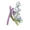 6ht5S S: Starting model for refinement |
|---|---|
| Similar structure data | |
| Other databases |
|
-Links
 PDBj
PDBj


- Assembly
| Deposited unit | 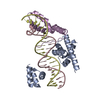
| ||||||||
|---|---|---|---|---|---|---|---|---|---|
| 1 |
| ||||||||
| Unit cell |
|
-Components
| #1: Protein | Mass: 10092.858 Da / Num. of mol.: 1 Source method: isolated from a genetically manipulated source Source: (gene. exp.)  |
|---|---|
| #2: DNA chain | Mass: 6752.419 Da / Num. of mol.: 1 Source method: isolated from a genetically manipulated source Source: (gene. exp.) |
| #3: DNA chain | Mass: 6747.374 Da / Num. of mol.: 1 Source method: isolated from a genetically manipulated source Source: (gene. exp.) |
| #4: Protein | Mass: 17962.875 Da / Num. of mol.: 1 Source method: isolated from a genetically manipulated source Source: (gene. exp.) |
| #5: Water | ChemComp-HOH /  Mass: 18.015 Da / Num. of mol.: 4 / Source method: isolated from a natural source / Formula: H2O Mass: 18.015 Da / Num. of mol.: 4 / Source method: isolated from a natural source / Formula: H2O |
-Experimental details
-Experiment
| Experiment | Method: X-RAY DIFFRACTION / Number of used crystals: 1 |
|---|
- Sample preparation
| Crystal | Density Matthews: 2.89 Å3/Da / Density % sol: 57.4 % |
|---|---|
| Crystal grow | Temperature: 277 K / Method: vapor diffusion, sitting drop / pH: 4.6 / Details: 0.1M sodium acetate, 12% (v/v) PEG 4000 |
-Data collection
| Diffraction | Mean temperature: 100 K / Serial crystal experiment: N |
|---|---|
| Diffraction source | Source: SYNCHROTRON / Site: PETRA III, EMBL c/o DESY  / Beamline: P13 (MX1) / Wavelength: 0.97624 Å / Beamline: P13 (MX1) / Wavelength: 0.97624 Å |
| Detector | Type: DECTRIS EIGER X 16M / Detector: PIXEL / Date: Jun 8, 2021 |
| Radiation | Monochromator: SI(111) / Protocol: SINGLE WAVELENGTH / Monochromatic (M) / Laue (L): M / Scattering type: x-ray |
| Radiation wavelength | Wavelength: 0.97624 Å / Relative weight: 1 |
| Reflection | Resolution: 2.5→126.92 Å / Num. obs: 15480 / % possible obs: 99.7 % / Redundancy: 11.4 % / CC1/2: 0.999 / Rmerge(I) obs: 0.091 / Net I/σ(I): 16.5 |
| Reflection shell | Resolution: 2.5→2.6 Å / Rmerge(I) obs: 1.28 / Mean I/σ(I) obs: 2 / Num. unique obs: 1711 / CC1/2: 0.755 |
- Processing
| Software |
| ||||||||||||||||||||||||||||||||||||||||||||||||||||||||||||||||||||||||||||||||||||||||||||||||||||||||||||||||||||||||||||||||||||||||||||||||||||||||||||||||||||||||||||||||||||||
|---|---|---|---|---|---|---|---|---|---|---|---|---|---|---|---|---|---|---|---|---|---|---|---|---|---|---|---|---|---|---|---|---|---|---|---|---|---|---|---|---|---|---|---|---|---|---|---|---|---|---|---|---|---|---|---|---|---|---|---|---|---|---|---|---|---|---|---|---|---|---|---|---|---|---|---|---|---|---|---|---|---|---|---|---|---|---|---|---|---|---|---|---|---|---|---|---|---|---|---|---|---|---|---|---|---|---|---|---|---|---|---|---|---|---|---|---|---|---|---|---|---|---|---|---|---|---|---|---|---|---|---|---|---|---|---|---|---|---|---|---|---|---|---|---|---|---|---|---|---|---|---|---|---|---|---|---|---|---|---|---|---|---|---|---|---|---|---|---|---|---|---|---|---|---|---|---|---|---|---|---|---|---|---|
| Refinement | Method to determine structure: MOLECULAR REPLACEMENT Starting model: 6ht5 Resolution: 2.5→58.48 Å / Cor.coef. Fo:Fc: 0.927 / Cor.coef. Fo:Fc free: 0.916 / SU B: 26.716 / SU ML: 0.255 / Cross valid method: THROUGHOUT / ESU R: 0.482 / ESU R Free: 0.29 / Stereochemistry target values: MAXIMUM LIKELIHOOD
| ||||||||||||||||||||||||||||||||||||||||||||||||||||||||||||||||||||||||||||||||||||||||||||||||||||||||||||||||||||||||||||||||||||||||||||||||||||||||||||||||||||||||||||||||||||||
| Solvent computation | Ion probe radii: 0.8 Å / Shrinkage radii: 0.8 Å / VDW probe radii: 1.2 Å / Solvent model: MASK | ||||||||||||||||||||||||||||||||||||||||||||||||||||||||||||||||||||||||||||||||||||||||||||||||||||||||||||||||||||||||||||||||||||||||||||||||||||||||||||||||||||||||||||||||||||||
| Displacement parameters | Biso mean: 75.215 Å2
| ||||||||||||||||||||||||||||||||||||||||||||||||||||||||||||||||||||||||||||||||||||||||||||||||||||||||||||||||||||||||||||||||||||||||||||||||||||||||||||||||||||||||||||||||||||||
| Refinement step | Cycle: 1 / Resolution: 2.5→58.48 Å
| ||||||||||||||||||||||||||||||||||||||||||||||||||||||||||||||||||||||||||||||||||||||||||||||||||||||||||||||||||||||||||||||||||||||||||||||||||||||||||||||||||||||||||||||||||||||
| Refine LS restraints |
|