 Movie
Movie Controller

[English] 日本語
 Yorodumi
- PDB-8bva: Crystal Structure of Mus musculus Protein Arginine Methyltransfer... -
+ Open data

- Basic information
| Entry | Database: PDB / ID: 8bva | ||||||
|---|---|---|---|---|---|---|---|
| Title | Crystal Structure of Mus musculus Protein Arginine Methyltransferase 2 in complex with RSF1_114-126 | ||||||
 Components Components |
| ||||||
 Keywords Keywords | TRANSFERASE / protein arginine N-methyltransferase / PRMT / SH3 / methylation / TRANSCRIPTION | ||||||
| Function / homology |  Function and homology information Function and homology informationtype I protein arginine methyltransferase / protein-arginine omega-N asymmetric methyltransferase activity / histone methyltransferase activity / mRNA splicing, via spliceosome / methylation / nuclear speck / mRNA binding / metal ion binding / nucleus / cytoplasm Similarity search - Function | ||||||
| Biological species |   Spodoptera aff. frugiperda 2 BOLD-2017 (butterflies/moths) Spodoptera aff. frugiperda 2 BOLD-2017 (butterflies/moths) | ||||||
| Method |  X-RAY DIFFRACTION / SYNCHROTRON / MOLECULAR REPLACEMENT / Resolution: 2.19 Å X-RAY DIFFRACTION / SYNCHROTRON / MOLECULAR REPLACEMENT / Resolution: 2.19 Å | ||||||
 Authors Authors | Cura, V. / Troffer-Charlier, N. / Marechal, N. / Bonnefond, L. / Cavarelli, J. | ||||||
| Funding support | 1items
| ||||||
 Citation Citation | Journal: To Be Published Title: Crystal Structure of Mus musculus Protein Arginine Methyltransferase 2 in complex with RSF1_114-126 Authors: Cura, V. / Troffer-Charlier, N. / Marechal, N. / Bonnefond, L. / Cavarelli, J. | ||||||
| History |
|
- Structure visualization
| Structure viewer | Molecule: MolmilJmol/JSmol |
|---|
- Downloads & links
-Download
| PDBx/mmCIF format | 8bva.cif.gz | 257.1 KB | Display | PDBx/mmCIF format |
|---|---|---|---|---|
| PDB format | pdb8bva.ent.gz | 173.4 KB | Display | PDB format |
| PDBx/mmJSON format | 8bva.json.gz | Tree view | PDBx/mmJSON format | |
| Others |  Other downloads Other downloads |
-Validation report
| Arichive directory | https://data.pdbj.org/pub/pdb/validation_reports/bv/8bvaftp://data.pdbj.org/pub/pdb/validation_reports/bv/8bva | HTTPS FTP |
|---|
-Related structure data
| Related structure data | 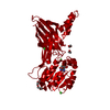 5fulS S: Starting model for refinement |
|---|---|
| Similar structure data |
-Links
 PDBj
PDBj
- Assembly
| Deposited unit | 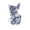
| ||||||||||
|---|---|---|---|---|---|---|---|---|---|---|---|
| 1 | 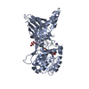
| ||||||||||
| Unit cell |
| ||||||||||
| Components on special symmetry positions |
|
-Components
-Protein / Protein/peptide , 2 types, 2 molecules AB
| #1: Protein | Mass: 39067.609 Da / Num. of mol.: 1 Source method: isolated from a genetically manipulated source Source: (gene. exp.) Production host: Spodoptera aff. frugiperda 1 BOLD-2017 (butterflies/moths)References: UniProt: Q3UKX1 |
|---|---|
| #2: Protein/peptide | Mass: 1470.510 Da / Num. of mol.: 1 / Source method: obtained synthetically Source: (synth.) Spodoptera aff. frugiperda 2 BOLD-2017 (butterflies/moths)References: UniProt: A0A2H1V327 |
-Non-polymers , 4 types, 88 molecules 


| #3: Chemical | ChemComp-TLA /  Mass: 150.087 Da / Num. of mol.: 1 / Source method: obtained synthetically / Formula: C4H6O6 Mass: 150.087 Da / Num. of mol.: 1 / Source method: obtained synthetically / Formula: C4H6O6 |
|---|---|
| #4: Chemical | ChemComp-K /  Mass: 39.098 Da / Num. of mol.: 1 / Source method: isolated from a natural source / Formula: K Mass: 39.098 Da / Num. of mol.: 1 / Source method: isolated from a natural source / Formula: K |
| #5: Chemical | ChemComp-QVR / ( Mass: 277.279 Da / Num. of mol.: 1 / Source method: obtained synthetically / Formula: C12H15N5O3 Mass: 277.279 Da / Num. of mol.: 1 / Source method: obtained synthetically / Formula: C12H15N5O3Source: (synth.) Spodoptera aff. frugiperda 2 BOLD-2017 (butterflies/moths)Feature type: SUBJECT OF INVESTIGATION |
| #6: Water | ChemComp-HOH / Mass: 18.015 Da / Num. of mol.: 85 / Source method: isolated from a natural source / Formula: H2O |
-Details
| Has ligand of interest | Y |
|---|---|
| Has protein modification | Y |
-Experimental details
-Experiment
| Experiment | Method: X-RAY DIFFRACTION / Number of used crystals: 1 |
|---|
- Sample preparation
| Crystal | Density Matthews: 3.05 Å3/Da / Density % sol: 59.72 % |
|---|---|
| Crystal grow | Temperature: 293 K / Method: vapor diffusion, hanging drop / Details: 1.2M ammonium tartrate |
-Data collection
| Diffraction | Mean temperature: 100 K / Serial crystal experiment: N |
|---|---|
| Diffraction source | Source: SYNCHROTRON / Site: ESRF  / Beamline: MASSIF-3 / Wavelength: 0.9677 Å / Beamline: MASSIF-3 / Wavelength: 0.9677 Å |
| Detector | Type: DECTRIS EIGER X 4M / Detector: PIXEL / Date: May 4, 2021 |
| Radiation | Protocol: SINGLE WAVELENGTH / Monochromatic (M) / Laue (L): M / Scattering type: x-ray |
| Radiation wavelength | Wavelength: 0.9677 Å / Relative weight: 1 |
| Reflection | Resolution: 2.19→29.55 Å / Num. obs: 25946 / % possible obs: 99.1 % / Redundancy: 13.8 % / Biso Wilson estimate: 44.5 Å2 / CC1/2: 0.999 / Rmerge(I) obs: 0.106 / Rrim(I) all: 0.11 / Χ2: 1 / Net I/σ(I): 14.9 |
| Reflection shell | Resolution: 2.19→2.26 Å / Redundancy: 12.3 % / Rmerge(I) obs: 1.48 / Mean I/σ(I) obs: 1.5 / Num. unique obs: 2056 / CC1/2: 0.686 / Rrim(I) all: 1.541 / Χ2: 0.97 / % possible all: 92.4 |
- Processing
| Software |
| ||||||||||||||||||||||||||||||||||||||||||||||||||||||||||||||||||||||||||||||||||||||||||||||||||||||||||||||||||||||||||||||||||||||||||||||||||||||
|---|---|---|---|---|---|---|---|---|---|---|---|---|---|---|---|---|---|---|---|---|---|---|---|---|---|---|---|---|---|---|---|---|---|---|---|---|---|---|---|---|---|---|---|---|---|---|---|---|---|---|---|---|---|---|---|---|---|---|---|---|---|---|---|---|---|---|---|---|---|---|---|---|---|---|---|---|---|---|---|---|---|---|---|---|---|---|---|---|---|---|---|---|---|---|---|---|---|---|---|---|---|---|---|---|---|---|---|---|---|---|---|---|---|---|---|---|---|---|---|---|---|---|---|---|---|---|---|---|---|---|---|---|---|---|---|---|---|---|---|---|---|---|---|---|---|---|---|---|---|---|---|
| Refinement | Method to determine structure: MOLECULAR REPLACEMENT Starting model: 5FUL Resolution: 2.19→29.55 Å / SU ML: 0.2088 / Cross valid method: FREE R-VALUE / σ(F): 1.35 / Phase error: 23.124 Stereochemistry target values: GeoStd + Monomer Library + CDL v1.2
| ||||||||||||||||||||||||||||||||||||||||||||||||||||||||||||||||||||||||||||||||||||||||||||||||||||||||||||||||||||||||||||||||||||||||||||||||||||||
| Solvent computation | Shrinkage radii: 0.9 Å / VDW probe radii: 1.11 Å / Solvent model: FLAT BULK SOLVENT MODEL | ||||||||||||||||||||||||||||||||||||||||||||||||||||||||||||||||||||||||||||||||||||||||||||||||||||||||||||||||||||||||||||||||||||||||||||||||||||||
| Displacement parameters | Biso mean: 54.24 Å2 | ||||||||||||||||||||||||||||||||||||||||||||||||||||||||||||||||||||||||||||||||||||||||||||||||||||||||||||||||||||||||||||||||||||||||||||||||||||||
| Refinement step | Cycle: LAST / Resolution: 2.19→29.55 Å
| ||||||||||||||||||||||||||||||||||||||||||||||||||||||||||||||||||||||||||||||||||||||||||||||||||||||||||||||||||||||||||||||||||||||||||||||||||||||
| Refine LS restraints |
| ||||||||||||||||||||||||||||||||||||||||||||||||||||||||||||||||||||||||||||||||||||||||||||||||||||||||||||||||||||||||||||||||||||||||||||||||||||||
| LS refinement shell |
| ||||||||||||||||||||||||||||||||||||||||||||||||||||||||||||||||||||||||||||||||||||||||||||||||||||||||||||||||||||||||||||||||||||||||||||||||||||||
| Refinement TLS params. | Method: refined / Refine-ID: X-RAY DIFFRACTION
| ||||||||||||||||||||||||||||||||||||||||||||||||||||||||||||||||||||||||||||||||||||||||||||||||||||||||||||||||||||||||||||||||||||||||||||||||||||||
| Refinement TLS group | Refine-ID: X-RAY DIFFRACTION
|
