 Movie
Movie Controller


+ Open data

- Basic information
| Entry | Database: PDB / ID: 8bv3 | ||||||
|---|---|---|---|---|---|---|---|
| Title | Bacillus subtilis DnaA domain III structure | ||||||
 Components Components | Chromosomal replication initiator protein DnaA | ||||||
 Keywords Keywords | CELL CYCLE / DnaA / DNA replication / DNA replication initiation / AAA+ superfamily / Initiator Clade | ||||||
| Function / homology |  Function and homology information Function and homology informationDnaA-oriC complex / nucleoid / regulation of DNA replication / DNA replication origin binding / DNA replication initiation / Hydrolases; Acting on acid anhydrides; Acting on acid anhydrides to facilitate cellular and subcellular movement / DNA replication / hydrolase activity / lipid binding / DNA-templated transcription ...DnaA-oriC complex / nucleoid / regulation of DNA replication / DNA replication origin binding / DNA replication initiation / Hydrolases; Acting on acid anhydrides; Acting on acid anhydrides to facilitate cellular and subcellular movement / DNA replication / hydrolase activity / lipid binding / DNA-templated transcription / ATP binding / identical protein binding / plasma membrane / cytoplasm Similarity search - Function | ||||||
| Biological species |  | ||||||
| Method |  X-RAY DIFFRACTION / SYNCHROTRON / MOLECULAR REPLACEMENT / Resolution: 2.38 Å X-RAY DIFFRACTION / SYNCHROTRON / MOLECULAR REPLACEMENT / Resolution: 2.38 Å | ||||||
 Authors Authors | Pintar, S. / Hubbard, J.A. | ||||||
| Funding support |  United Kingdom, 1items United Kingdom, 1items
| ||||||
 Citation Citation | Journal: Nat Commun / Year: 2023 Title: The bacterial replication origin BUS promotes nucleobase capture. Authors: Simone Pelliciari / Salomé Bodet-Lefèvre / Stepan Fenyk / Daniel Stevens / Charles Winterhalter / Frederic D Schramm / Sara Pintar / Daniel R Burnham / George Merces / Tomas T Richardson / ...Authors: Simone Pelliciari / Salomé Bodet-Lefèvre / Stepan Fenyk / Daniel Stevens / Charles Winterhalter / Frederic D Schramm / Sara Pintar / Daniel R Burnham / George Merces / Tomas T Richardson / Yumiko Tashiro / Julia Hubbard / Hasan Yardimci / Aravindan Ilangovan / Heath Murray / Abstract: Genome duplication is essential for the proliferation of cellular life and this process is generally initiated by dedicated replication proteins at chromosome origins. In bacteria, DNA replication is ...Genome duplication is essential for the proliferation of cellular life and this process is generally initiated by dedicated replication proteins at chromosome origins. In bacteria, DNA replication is initiated by the ubiquitous DnaA protein, which assembles into an oligomeric complex at the chromosome origin (oriC) that engages both double-stranded DNA (dsDNA) and single-stranded DNA (ssDNA) to promote DNA duplex opening. However, the mechanism of DnaA specifically opening a replication origin was unknown. Here we show that Bacillus subtilis DnaA assembles into a continuous oligomer at the site of DNA melting, extending from a dsDNA anchor to engage a single DNA strand. Within this complex, two nucleobases of each ssDNA binding motif (DnaA-trio) are captured within a dinucleotide binding pocket created by adjacent DnaA proteins. These results provide a molecular basis for DnaA specifically engaging the conserved sequence elements within the bacterial chromosome origin basal unwinding system (BUS). | ||||||
| History |
|
- Structure visualization
| Structure viewer | Molecule: MolmilJmol/JSmol |
|---|
- Downloads & links
-Download
| PDBx/mmCIF format | 8bv3.cif.gz | 244.7 KB | Display | PDBx/mmCIF format |
|---|---|---|---|---|
| PDB format | pdb8bv3.ent.gz | 197.8 KB | Display | PDB format |
| PDBx/mmJSON format | 8bv3.json.gz | Tree view | PDBx/mmJSON format | |
| Others |  Other downloads Other downloads |
-Validation report
| Arichive directory | https://data.pdbj.org/pub/pdb/validation_reports/bv/8bv3ftp://data.pdbj.org/pub/pdb/validation_reports/bv/8bv3 | HTTPS FTP |
|---|
-Related structure data
| Related structure data | 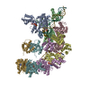 8btgC 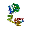 1l8qS S: Starting model for refinement C: citing same article ( |
|---|---|
| Similar structure data |


-Links
 PDBj
PDBj


- Assembly
| Deposited unit | 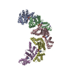
| ||||||||
|---|---|---|---|---|---|---|---|---|---|
| 1 |
| ||||||||
| Unit cell |
|
-Components
| #1: Protein | Mass: 27466.150 Da / Num. of mol.: 5 Source method: isolated from a genetically manipulated source Source: (gene. exp.) #2: Chemical | ChemComp-ANP /   Mass: 506.196 Da / Num. of mol.: 5 / Source method: obtained synthetically / Formula: C10H17N6O12P3 / Feature type: SUBJECT OF INVESTIGATION / Comment: AMP-PNP, energy-carrying molecule analogue*YM Mass: 506.196 Da / Num. of mol.: 5 / Source method: obtained synthetically / Formula: C10H17N6O12P3 / Feature type: SUBJECT OF INVESTIGATION / Comment: AMP-PNP, energy-carrying molecule analogue*YM#3: Chemical | ChemComp-K /   Mass: 39.098 Da / Num. of mol.: 9 / Source method: obtained synthetically / Formula: K Mass: 39.098 Da / Num. of mol.: 9 / Source method: obtained synthetically / Formula: K#4: Chemical | ChemComp-MG /   Mass: 24.305 Da / Num. of mol.: 5 / Source method: obtained synthetically / Formula: Mg / Feature type: SUBJECT OF INVESTIGATION Mass: 24.305 Da / Num. of mol.: 5 / Source method: obtained synthetically / Formula: Mg / Feature type: SUBJECT OF INVESTIGATION#5: Water | ChemComp-HOH / |  Mass: 18.015 Da / Num. of mol.: 122 / Source method: isolated from a natural source / Formula: H2O Mass: 18.015 Da / Num. of mol.: 122 / Source method: isolated from a natural source / Formula: H2OHas ligand of interest | Y | Has protein modification | N | |
|---|
-Experimental details
-Experiment
| Experiment | Method: X-RAY DIFFRACTION / Number of used crystals: 1 |
|---|
- Sample preparation
| Crystal | Density Matthews: 3.12 Å3/Da / Density % sol: 60.61 % |
|---|---|
| Crystal grow | Temperature: 293.15 K / Method: evaporation Details: Morpheus H3: 0.02 M DL-Glutamic acid monohydrate, 0.02 M DL-Alanine, 0.0 2M Glycine, 0.2M DL-Lysine monohydrochloride, 0.02M DL-Serine, 0.1 M Imidazole/ MES monohydrate pH 6.5, 20% v/v Glycerol, 20% w/v PEG 4000 |
-Data collection
| Diffraction | Mean temperature: 100 K / Serial crystal experiment: N |
|---|---|
| Diffraction source | Source: SYNCHROTRON / Site: Diamond / Beamline: I24 / Wavelength: 0.99987 Å |
| Detector | Type: DECTRIS PILATUS3 6M / Detector: PIXEL / Date: Feb 25, 2022 |
| Radiation | Protocol: SINGLE WAVELENGTH / Monochromatic (M) / Laue (L): M / Scattering type: x-ray |
| Radiation wavelength | Wavelength: 0.99987 Å / Relative weight: 1 |
| Reflection | Resolution: 2.38→74.399 Å / Num. obs: 56743 / % possible obs: 85.3 % / Redundancy: 9.7 % / CC1/2: 0.996 / Rmerge(I) obs: 0.179 / Rpim(I) all: 0.06 / Rrim(I) all: 0.189 / Net I/σ(I): 10.1 |
| Reflection shell | Resolution: 2.38→2.52 Å / Redundancy: 6.7 % / Rmerge(I) obs: 1.461 / Mean I/σ(I) obs: 1.5 / Num. unique obs: 2842 / CC1/2: 0.461 / Rpim(I) all: 0.599 / Rrim(I) all: 1.583 / % possible all: 27.2 |
- Processing
| Software |
| ||||||||||||||||||||||||
|---|---|---|---|---|---|---|---|---|---|---|---|---|---|---|---|---|---|---|---|---|---|---|---|---|---|
| Refinement | Method to determine structure: MOLECULAR REPLACEMENT Starting model: 1L8Q Resolution: 2.38→74.399 Å / Cor.coef. Fo:Fc: 0.952 / Cor.coef. Fo:Fc free: 0.924 / SU B: 0.004 / SU ML: 0 / Cross valid method: THROUGHOUT / σ(F): 0 / ESU R: 0.206 / ESU R Free: 0.258 / Stereochemistry target values: MAXIMUM LIKELIHOOD Details: HYDROGENS HAVE BEEN ADDED IN THE RIDING POSITIONS U VALUES : REFINED INDIVIDUALLY
| ||||||||||||||||||||||||
| Solvent computation | Ion probe radii: 0.8 Å / Shrinkage radii: 0.8 Å / VDW probe radii: 1.2 Å / Solvent model: MASK | ||||||||||||||||||||||||
| Displacement parameters | Biso max: 117.19 Å2 / Biso mean: 50.058 Å2 / Biso min: 19.32 Å2
| ||||||||||||||||||||||||
| Refinement step | Cycle: final / Resolution: 2.38→74.399 Å
| ||||||||||||||||||||||||
| LS refinement shell | Resolution: 2.381→2.443 Å / Rfactor Rfree error: 0 / Total num. of bins used: 20
|
