Entry Database : PDB / ID : 8bseTitle CRYSTAL STRUCTURE OF SARS-COV-2 RECEPTOR BINDING DOMAIN (RBD) in complex with 1D1 Fab 1D1 FAB HEAVY CHAIN 1D1 FAB LIGHT CHAIN Spike glycoprotein Keywords / / / / / Function / homology Function Domain/homology Component
/ / / / / / / / / / / / / / / / / / / / / / / / / / / / / / / / / / / / / / / / / / / / / / / / / / / / / / / / / / / / / / / / Biological species Homo sapiens (human)Method / / / Resolution : 1.9 Å Authors Welin, M. / Kimbung, Y.R. / Focht, D. / Pisitkun, T. Funding support Organization Grant number Country Other government
Journal : Plos One / Year : 2023Title : Efficacy of the combination of monoclonal antibodies against the SARS-CoV-2 Beta and Delta variants.Authors: Boonkrai, C. / Cotrone, T.S. / Chaisuriyong, W. / Tantawichien, T. / Thisyakorn, U. / Fernandez, S. / Hunsawong, T. / Reed, M. / Wongtangprasert, T. / Audomsun, T. / Phakham, T. / ... Authors : Boonkrai, C. / Cotrone, T.S. / Chaisuriyong, W. / Tantawichien, T. / Thisyakorn, U. / Fernandez, S. / Hunsawong, T. / Reed, M. / Wongtangprasert, T. / Audomsun, T. / Phakham, T. / Attakitbancha, C. / Saelao, P. / Focht, D. / Kimbung, R. / Welin, M. / Malik, A.A. / Pisitkun, T. / Srisawat, N. History Deposition Nov 25, 2022 Deposition site / Processing site Revision 1.0 May 17, 2023 Provider / Type Revision 1.1 Oct 18, 2023 Group / Database references / Source and taxonomyCategory chem_comp_atom / chem_comp_bond ... chem_comp_atom / chem_comp_bond / citation / citation_author / entity_src_gen Item _citation.country / _citation.journal_abbrev ... _citation.country / _citation.journal_abbrev / _citation.journal_id_CSD / _citation.journal_id_ISSN / _citation.journal_volume / _citation.page_first / _citation.page_last / _citation.pdbx_database_id_DOI / _citation.pdbx_database_id_PubMed / _citation.title / _citation.year / _entity_src_gen.pdbx_host_org_ncbi_taxonomy_id / _entity_src_gen.pdbx_host_org_scientific_name
Show all Show less
 Movie
Movie Controller

 Yorodumi
 Open data
 Basic information
 Components
 Keywords
 Function and homology information
 Severe acute respiratory syndrome coronavirus 2
 Homo sapiens (human)
 X-RAY DIFFRACTION /
  Authors
 Thailand, 1items
  Citation
 Structure visualization
 Downloads & links
 Other downloads
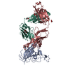
 PDBj
PDBj


 Assembly
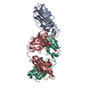

 Trichoplusia ni (cabbage looper) / References: UniProt: P0DTC2
 Type: D-saccharide, beta linking / Mass: 221.208 Da / Num. of mol.: 1 / Source method: obtained synthetically / Formula: C8H15NO6


 Mass: 92.094 Da / Num. of mol.: 5 / Source method: obtained synthetically / Formula: C3H8O3
 Mass: 122.143 Da / Num. of mol.: 1 / Source method: obtained synthetically / Formula: C4H12NO3 / Comment: pH buffer*YM
 Sample preparation
 / Beamline: BioMAX / Wavelength: 0.9537 Å
 Processing