 Movie
Movie Controller


+ Open data

- Basic information
| Entry | Database: PDB / ID: 8bqf | |||||||||
|---|---|---|---|---|---|---|---|---|---|---|
| Title | Adenylate Kinase L107I MUTANT | |||||||||
 Components Components | Adenylate kinase | |||||||||
 Keywords Keywords | TRANSFERASE / COMPLEX | |||||||||
| Function / homology |  Function and homology information Function and homology informationpurine ribonucleotide interconversion / adenine metabolic process / nucleoside monophosphate metabolic process / ADP biosynthetic process / nucleoside diphosphate metabolic process / adenylate kinase / AMP kinase activity / AMP salvage / nucleoside diphosphate kinase activity / AMP binding ...purine ribonucleotide interconversion / adenine metabolic process / nucleoside monophosphate metabolic process / ADP biosynthetic process / nucleoside diphosphate metabolic process / adenylate kinase / AMP kinase activity / AMP salvage / nucleoside diphosphate kinase activity / AMP binding / magnesium ion binding / ATP binding / cytoplasm / cytosol Similarity search - Function | |||||||||
| Biological species |  | |||||||||
| Method |  X-RAY DIFFRACTION / MOLECULAR REPLACEMENT / Resolution: 2.05 Å X-RAY DIFFRACTION / MOLECULAR REPLACEMENT / Resolution: 2.05 Å | |||||||||
 Authors Authors | Scheerer, D. / Adkar, B.V. / Bhattacharyya, S. / Levy, D. / Iljina, M. / Iljina, I. / Dym, O. / Haran, G. / Shakhnovich, E.I. | |||||||||
| Funding support | European Union,  Israel, 2items Israel, 2items
| |||||||||
 Citation Citation | Journal: Proc.Natl.Acad.Sci.USA / Year: 2023 Title: Allosteric communication between ligand binding domains modulates substrate inhibition in adenylate kinase. Authors: Scheerer, D. / Adkar, B.V. / Bhattacharyya, S. / Levy, D. / Iljina, M. / Riven, I. / Dym, O. / Haran, G. / Shakhnovich, E.I. | |||||||||
| History |
|
- Structure visualization
| Structure viewer | Molecule: MolmilJmol/JSmol |
|---|
- Downloads & links
-Download
| PDBx/mmCIF format | 8bqf.cif.gz | 257.4 KB | Display | PDBx/mmCIF format |
|---|---|---|---|---|
| PDB format | pdb8bqf.ent.gz | 206.8 KB | Display | PDB format |
| PDBx/mmJSON format | 8bqf.json.gz | Tree view | PDBx/mmJSON format | |
| Others |  Other downloads Other downloads |
-Validation report
| Arichive directory | https://data.pdbj.org/pub/pdb/validation_reports/bq/8bqfftp://data.pdbj.org/pub/pdb/validation_reports/bq/8bqf | HTTPS FTP |
|---|
-Related structure data
| Related structure data |  7f7u S: Starting model for refinement |
|---|---|
| Similar structure data |
-Links
 PDBj
PDBj- Assembly
| Deposited unit | 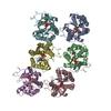
| ||||||||
|---|---|---|---|---|---|---|---|---|---|
| 1 | 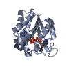
| ||||||||
| 2 | 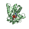
| ||||||||
| 3 | 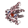
| ||||||||
| 4 | 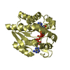
| ||||||||
| 5 | 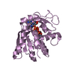
| ||||||||
| 6 | 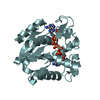
| ||||||||
| Unit cell |
|
-Components
| #1: Protein | Mass: 25791.402 Da / Num. of mol.: 6 / Mutation: L107I Source method: isolated from a genetically manipulated source Source: (gene. exp.) #2: Chemical | ChemComp-AP5 /   Mass: 916.367 Da / Num. of mol.: 6 / Source method: obtained synthetically / Formula: C20H29N10O22P5 / Feature type: SUBJECT OF INVESTIGATION Mass: 916.367 Da / Num. of mol.: 6 / Source method: obtained synthetically / Formula: C20H29N10O22P5 / Feature type: SUBJECT OF INVESTIGATION#3: Water | ChemComp-HOH / |  Mass: 18.015 Da / Num. of mol.: 223 / Source method: isolated from a natural source / Formula: H2O Mass: 18.015 Da / Num. of mol.: 223 / Source method: isolated from a natural source / Formula: H2OHas ligand of interest | Y | Has protein modification | N | |
|---|
-Experimental details
-Experiment
| Experiment | Method: X-RAY DIFFRACTION / Number of used crystals: 1 |
|---|
- Sample preparation
| Crystal | Density Matthews: 2.33 Å3/Da / Density % sol: 47.11 % |
|---|---|
| Crystal grow | Temperature: 292 K / Method: vapor diffusion, hanging drop / pH: 7 Details: 7.5% PEG 3,350, 7.5% PEG 4,000, 7.5% PEG 2,000, 7.5% PEG 5,000 monomethyl ether, 0.07M ammonium nitrate, 2.5% ethylene glycol and 0.05M MES pH=7. PH range: 7 |
-Data collection
| Diffraction | Mean temperature: 100 K / Serial crystal experiment: N |
|---|---|
| Diffraction source | Source: ROTATING ANODE / Type: RIGAKU / Wavelength: 1.3405 Å |
| Detector | Type: RIGAKU HyPix-3000 / Detector: PIXEL / Date: Mar 20, 2022 |
| Radiation | Monochromator: Ga / Protocol: SINGLE WAVELENGTH / Monochromatic (M) / Laue (L): M / Scattering type: x-ray |
| Radiation wavelength | Wavelength: 1.3405 Å / Relative weight: 1 |
| Reflection | Resolution: 2.05→21.04 Å / Num. obs: 93318 / % possible obs: 99.69 % / Redundancy: 8.1 % / CC1/2: 0.995 / CC star: 0.999 / Rmerge(I) obs: 0.1043 / Rpim(I) all: 0.03909 / Rrim(I) all: 0.1117 / Net I/σ(I): 20.47 |
| Reflection shell | Resolution: 2.05→2.123 Å / Redundancy: 7.4 % / Rmerge(I) obs: 0.4044 / Mean I/σ(I) obs: 4.39 / Num. unique obs: 8992 / CC1/2: 0.931 / CC star: 0.982 / Rpim(I) all: 0.1585 / Rrim(I) all: 0.435 / % possible all: 100 |
- Processing
| Software |
| ||||||||||||||||||||||||||||||||||||||||||||||||||||||||||||||||||||||||||||||||||||||||||||||||||||||||||||||||||||||||||||||||||||||||||||||||||||||||||||||||||||||||||||||||||||||
|---|---|---|---|---|---|---|---|---|---|---|---|---|---|---|---|---|---|---|---|---|---|---|---|---|---|---|---|---|---|---|---|---|---|---|---|---|---|---|---|---|---|---|---|---|---|---|---|---|---|---|---|---|---|---|---|---|---|---|---|---|---|---|---|---|---|---|---|---|---|---|---|---|---|---|---|---|---|---|---|---|---|---|---|---|---|---|---|---|---|---|---|---|---|---|---|---|---|---|---|---|---|---|---|---|---|---|---|---|---|---|---|---|---|---|---|---|---|---|---|---|---|---|---|---|---|---|---|---|---|---|---|---|---|---|---|---|---|---|---|---|---|---|---|---|---|---|---|---|---|---|---|---|---|---|---|---|---|---|---|---|---|---|---|---|---|---|---|---|---|---|---|---|---|---|---|---|---|---|---|---|---|---|---|
| Refinement | Method to determine structure: MOLECULAR REPLACEMENT Starting model: 7F7U 7f7u Resolution: 2.05→21.04 Å / Cor.coef. Fo:Fc: 0.938 / Cor.coef. Fo:Fc free: 0.919 / SU B: 4.648 / SU ML: 0.129 / Cross valid method: THROUGHOUT / ESU R: 0.194 / ESU R Free: 0.173 / Stereochemistry target values: MAXIMUM LIKELIHOOD / Details: HYDROGENS HAVE BEEN ADDED IN THE RIDING POSITIONS
| ||||||||||||||||||||||||||||||||||||||||||||||||||||||||||||||||||||||||||||||||||||||||||||||||||||||||||||||||||||||||||||||||||||||||||||||||||||||||||||||||||||||||||||||||||||||
| Solvent computation | Ion probe radii: 0.8 Å / Shrinkage radii: 0.8 Å / VDW probe radii: 1.2 Å / Solvent model: MASK | ||||||||||||||||||||||||||||||||||||||||||||||||||||||||||||||||||||||||||||||||||||||||||||||||||||||||||||||||||||||||||||||||||||||||||||||||||||||||||||||||||||||||||||||||||||||
| Displacement parameters | Biso mean: 27.461 Å2
| ||||||||||||||||||||||||||||||||||||||||||||||||||||||||||||||||||||||||||||||||||||||||||||||||||||||||||||||||||||||||||||||||||||||||||||||||||||||||||||||||||||||||||||||||||||||
| Refinement step | Cycle: 1 / Resolution: 2.05→21.04 Å
| ||||||||||||||||||||||||||||||||||||||||||||||||||||||||||||||||||||||||||||||||||||||||||||||||||||||||||||||||||||||||||||||||||||||||||||||||||||||||||||||||||||||||||||||||||||||
| Refine LS restraints |
|