登録構造単位
A: Disks large-like protein 1
B: Disks large-like protein 1
C: Disks large-like protein 1
D: Vang-like protein 1
F: Vang-like protein 1
E: Vang-like protein 1 概要 構成要素の詳細
分子量 (理論値) 分子数 合計 (水以外) 32,272 6 ポリマ- 32,272 6 非ポリマー 0 0 水 955 53
1 概要 対称操作
登録構造と同一 登録者が定義した集合体 根拠 :
タイプ 名称 対称操作 数 identity operation 1_555 x,y,z 1
単位格子 Length a, b, c (Å) 64.673, 101.980, 106.063 Angle α, β, γ (deg.) 90.000, 90.000, 90.000 Int Tables number 20 Space group name H-M C2221 Space group name Hall C2c2 Symmetry operation #1 : x,y,z#2 : x,-y,-z#3 : -x,y,-z+1/2#4 : -x,-y,z+1/2#5 : x+1/2,y+1/2,z#6 : x+1/2,-y+1/2,-z#7 : -x+1/2,y+1/2,-z+1/2#8 : -x+1/2,-y+1/2,z+1/2
非結晶学的対称性 (NCS) NCSドメイン ID Ens-ID 詳細 d_1ens_1(chain "A" and (resid 316 through 325 or (resid 326...d_2ens_1(chain "B" and (resid 316 through 360 or (resid 361...d_3ens_1(chain "C" and (resid 316 through 324 or (resid 325...d_1ens_2chain "D"d_2ens_2chain "F"
NCSドメイン領域 Dom-ID Component-ID Ens-ID Beg label comp-ID End label comp-ID Label asym-ID Label seq-ID d_11 ens_1SERPROA1 - 89 d_21 ens_1SERPROB2 - 90 d_31 ens_1SERASPC4 - 14 d_32 ens_1GLYPROC16 - 93 d_11 ens_2PROVALD1 - 5 d_21 ens_2PROVALE1 - 5
NCSアンサンブル NCS oper ID Code Matrix ベクター 1 given(-0.671590763585, 0.18141648297, -0.718368920524), (0.149530878418, -0.916423032504, -0.371226806543), (-0.72567648623, -0.356730830183, 0.588333878108)-0.607554830378, -48.9917741389, -11.50010094692 given(-0.445316281924, -0.647315153297, -0.618608520282), (0.527979774779, 0.368151749367, -0.765311470489), (0.723139520755, -0.667418445767, 0.177825340638)-27.0894198399, -19.7356594867, -30.8653481173 given(-0.520799615416, -0.624968609361, -0.581534176034), (0.528341794665, 0.299100667571, -0.79460288111), (0.670539117869, -0.721077685115, 0.174425524037)-26.6905411008, -20.4928613577, -31.1835695691
 ムービー
ムービー コントローラー


 データを開く
 基本情報
基本情報 要素
要素 キーワード
キーワード 機能・相同性情報
 X線回折 /
  データ登録者
 オーストラリア, 2件
  引用
引用 構造の表示
構造の表示 ダウンロードとリンク
 その他のダウンロード
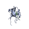
 PDBj
PDBj 集合体
集合体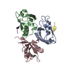

 分子量: 18.015 Da / 分子数: 53 / 由来タイプ: 天然 / 式: H2O
 試料調製
試料調製 解析
解析