 Movie
Movie Controller

[English] 日本語
 Yorodumi
- PDB-8bkn: Carboxymyoglobin dark state for comparison with 5 mJ/cm2 time series -
+ Open data

- Basic information
| Entry | Database: PDB / ID: 8bkn | ||||||
|---|---|---|---|---|---|---|---|
| Title | Carboxymyoglobin dark state for comparison with 5 mJ/cm2 time series | ||||||
 Components Components | Myoglobin | ||||||
 Keywords Keywords | OXYGEN STORAGE / XFEL / SFX / photolysis / pump-probe / Free-electron Laser / Serial Femtosecond Crystallography | ||||||
| Function / homology |  Function and homology information Function and homology informationOxidoreductases; Acting on other nitrogenous compounds as donors / nitrite reductase activity / oxygen transport / sarcoplasm / Oxidoreductases; Acting on a peroxide as acceptor; Peroxidases / removal of superoxide radicals / oxygen carrier activity / peroxidase activity / oxygen binding / heme binding / metal ion binding Similarity search - Function | ||||||
| Biological species |  | ||||||
| Method |  X-RAY DIFFRACTION / FREE ELECTRON LASER / MOLECULAR REPLACEMENT / Resolution: 1.29 Å X-RAY DIFFRACTION / FREE ELECTRON LASER / MOLECULAR REPLACEMENT / Resolution: 1.29 Å | ||||||
 Authors Authors | Barends, T. / Schlichting, I. | ||||||
| Funding support |  Germany, 1items Germany, 1items
| ||||||
 Citation Citation | Journal: Nature / Year: 2024 Title: Influence of pump laser fluence on ultrafast myoglobin structural dynamics. Authors: Barends, T.R.M. / Gorel, A. / Bhattacharyya, S. / Schiro, G. / Bacellar, C. / Cirelli, C. / Colletier, J.P. / Foucar, L. / Grunbein, M.L. / Hartmann, E. / Hilpert, M. / Holton, J.M. / ...Authors: Barends, T.R.M. / Gorel, A. / Bhattacharyya, S. / Schiro, G. / Bacellar, C. / Cirelli, C. / Colletier, J.P. / Foucar, L. / Grunbein, M.L. / Hartmann, E. / Hilpert, M. / Holton, J.M. / Johnson, P.J.M. / Kloos, M. / Knopp, G. / Marekha, B. / Nass, K. / Nass Kovacs, G. / Ozerov, D. / Stricker, M. / Weik, M. / Doak, R.B. / Shoeman, R.L. / Milne, C.J. / Huix-Rotllant, M. / Cammarata, M. / Schlichting, I. | ||||||
| History |
|
- Structure visualization
| Structure viewer | Molecule: MolmilJmol/JSmol |
|---|
- Downloads & links
-Download
| PDBx/mmCIF format | 8bkn.cif.gz | 49.8 KB | Display | PDBx/mmCIF format |
|---|---|---|---|---|
| PDB format | pdb8bkn.ent.gz | 32.8 KB | Display | PDB format |
| PDBx/mmJSON format | 8bkn.json.gz | Tree view | PDBx/mmJSON format | |
| Others |  Other downloads Other downloads |
-Validation report
| Arichive directory | https://data.pdbj.org/pub/pdb/validation_reports/bk/8bknftp://data.pdbj.org/pub/pdb/validation_reports/bk/8bkn | HTTPS FTP |
|---|
-Related structure data
| Related structure data |  8bkhC  8r8fC  8r8gC  8r8hC  8r8iC  8r8jC  8r8wC  8r8xC  8r8yC  8r8zC  8r90C  8r91C  8r92C  8r93C  8r94C  8r95C  8r9cC  8r9dC  8r9eC  8r9fC  8r9gC  8r9hC  8r9iC  8r9jC  8r9kC  8r9lC  8r9mC  8r9nC  8r9pC  8r9qC  8ra1C  8ra2C  8ra3C  8ra4C  8ra5C  8ra6C  8ra7C  8ra8C  8ra9C  8raaC  8rabC  8racC  8radC  8raeC 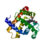 5d5rS S: Starting model for refinement C: citing same article ( |
|---|---|
| Similar structure data |
-Links
 PDBj
PDBj


- Assembly
| Deposited unit | 
| ||||||||||||
|---|---|---|---|---|---|---|---|---|---|---|---|---|---|
| 1 |
| ||||||||||||
| Unit cell |
|
-Components
| #1: Protein | Mass: 16926.463 Da / Num. of mol.: 1 / Source method: isolated from a natural source / Source: (natural) | ||||||
|---|---|---|---|---|---|---|---|
| #2: Chemical | ChemComp-HEM /   Mass: 616.487 Da / Num. of mol.: 1 / Source method: obtained synthetically / Formula: C34H32FeN4O4 / Feature type: SUBJECT OF INVESTIGATION Mass: 616.487 Da / Num. of mol.: 1 / Source method: obtained synthetically / Formula: C34H32FeN4O4 / Feature type: SUBJECT OF INVESTIGATION | ||||||
| #3: Chemical |   Mass: 96.063 Da / Num. of mol.: 2 / Source method: obtained synthetically / Formula: SO4 Mass: 96.063 Da / Num. of mol.: 2 / Source method: obtained synthetically / Formula: SO4#4: Chemical | ChemComp-CMO / |   Mass: 28.010 Da / Num. of mol.: 1 / Source method: obtained synthetically / Formula: CO Mass: 28.010 Da / Num. of mol.: 1 / Source method: obtained synthetically / Formula: CO#5: Water | ChemComp-HOH / |  Mass: 18.015 Da / Num. of mol.: 130 / Source method: isolated from a natural source / Formula: H2O Mass: 18.015 Da / Num. of mol.: 130 / Source method: isolated from a natural source / Formula: H2OHas ligand of interest | Y | |
-Experimental details
-Experiment
| Experiment | Method: X-RAY DIFFRACTION / Number of used crystals: 1 |
|---|
- Sample preparation
| Crystal | Density Matthews: 1.78 Å3/Da / Density % sol: 31.06 % |
|---|---|
| Crystal grow | Temperature: 293 K / Method: batch mode / Details: 3.1 M ammonium sulfate |
-Data collection
| Diffraction | Mean temperature: 293 K / Serial crystal experiment: N |
|---|---|
| Diffraction source | Source: FREE ELECTRON LASER / Type: OTHER / Wavelength: 1.03 Å |
| Detector | Type: CUSTOM-MADE / Detector: PIXEL / Date: May 1, 2019 |
| Radiation | Protocol: SINGLE WAVELENGTH / Monochromatic (M) / Laue (L): M / Scattering type: x-ray |
| Radiation wavelength | Wavelength: 1.03 Å / Relative weight: 1 |
| Reflection | Resolution: 1.29→30.12 Å / Num. obs: 30443 / % possible obs: 99.9 % / Redundancy: 264.9 % / Biso Wilson estimate: 13.86 Å2 / CC star: 0.992 / R split: 0.147 / Net I/σ(I): 5.7 |
| Reflection shell | Resolution: 1.29→1.32 Å / Num. unique obs: 1976 / CC star: 0.835 / R split: 0.658 |
- Processing
| Software | Name: PHENIX / Version: 1.19.2_4158 / Classification: refinement | |||||||||||||||||||||||||||||||||||||||||||||||||||||||||||||||||||||||||||||||||||||||||||||||||||||||||||||||||||||||
|---|---|---|---|---|---|---|---|---|---|---|---|---|---|---|---|---|---|---|---|---|---|---|---|---|---|---|---|---|---|---|---|---|---|---|---|---|---|---|---|---|---|---|---|---|---|---|---|---|---|---|---|---|---|---|---|---|---|---|---|---|---|---|---|---|---|---|---|---|---|---|---|---|---|---|---|---|---|---|---|---|---|---|---|---|---|---|---|---|---|---|---|---|---|---|---|---|---|---|---|---|---|---|---|---|---|---|---|---|---|---|---|---|---|---|---|---|---|---|---|---|
| Refinement | Method to determine structure: MOLECULAR REPLACEMENT Starting model: 5d5r Resolution: 1.29→30.12 Å / SU ML: 0.1732 / Cross valid method: FREE R-VALUE / σ(F): 1.35 / Phase error: 20.8649 Stereochemistry target values: GeoStd + Monomer Library + CDL v1.2
| |||||||||||||||||||||||||||||||||||||||||||||||||||||||||||||||||||||||||||||||||||||||||||||||||||||||||||||||||||||||
| Solvent computation | Shrinkage radii: 0.9 Å / VDW probe radii: 1.11 Å / Solvent model: FLAT BULK SOLVENT MODEL | |||||||||||||||||||||||||||||||||||||||||||||||||||||||||||||||||||||||||||||||||||||||||||||||||||||||||||||||||||||||
| Displacement parameters | Biso mean: 17.6 Å2 | |||||||||||||||||||||||||||||||||||||||||||||||||||||||||||||||||||||||||||||||||||||||||||||||||||||||||||||||||||||||
| Refinement step | Cycle: LAST / Resolution: 1.29→30.12 Å
| |||||||||||||||||||||||||||||||||||||||||||||||||||||||||||||||||||||||||||||||||||||||||||||||||||||||||||||||||||||||
| Refine LS restraints |
| |||||||||||||||||||||||||||||||||||||||||||||||||||||||||||||||||||||||||||||||||||||||||||||||||||||||||||||||||||||||
| LS refinement shell |
|
