Deposited unit
A: Mutant Imine Reductase IR007-143 from Amycolatopsis azurea, E120A, M197W, M206S, A213P, D238G, I240L
B: Mutant Imine Reductase IR007-143 from Amycolatopsis azurea, E120A, M197W, M206S, A213P, D238G, I240L
C: Mutant Imine Reductase IR007-143 from Amycolatopsis azurea, E120A, M197W, M206S, A213P, D238G, I240L
D: Mutant Imine Reductase IR007-143 from Amycolatopsis azurea, E120A, M197W, M206S, A213P, D238G, I240L
hetero molecules Summary Component details
Theoretical mass Number of molelcules Total (without water) 127,394 16 Polymers 123,652 4 Non-polymers 3,742 12 Water 4,179 232
1
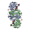
A: Mutant Imine Reductase IR007-143 from Amycolatopsis azurea, E120A, M197W, M206S, A213P, D238G, I240L
B: Mutant Imine Reductase IR007-143 from Amycolatopsis azurea, E120A, M197W, M206S, A213P, D238G, I240L
hetero molecules Summary Component details Symmetry operations Calculated values
Theoretical mass Number of molelcules Total (without water) 63,793 9 Polymers 61,826 2 Non-polymers 1,967 7 Water 36 2
Type Name Symmetry operation Number identity operation 1_555 x,y,z 1
Buried area 12430 Å2 ΔGint -138 kcal/mol Surface area 21070 Å2 Method
2
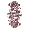
C: Mutant Imine Reductase IR007-143 from Amycolatopsis azurea, E120A, M197W, M206S, A213P, D238G, I240L
hetero molecules
C: Mutant Imine Reductase IR007-143 from Amycolatopsis azurea, E120A, M197W, M206S, A213P, D238G, I240L
hetero molecules Summary Component details Symmetry operations Calculated values
Theoretical mass Number of molelcules Total (without water) 63,697 8 Polymers 61,826 2 Non-polymers 1,871 6 Water 36 2
Type Name Symmetry operation Number identity operation 1_555 x,y,z 1 crystal symmetry operation 17_554 x-y+1/3,-y+2/3,-z-1/3 1
Buried area 12350 Å2 ΔGint -125 kcal/mol Surface area 20950 Å2 Method
3
D: Mutant Imine Reductase IR007-143 from Amycolatopsis azurea, E120A, M197W, M206S, A213P, D238G, I240L
hetero molecules
D: Mutant Imine Reductase IR007-143 from Amycolatopsis azurea, E120A, M197W, M206S, A213P, D238G, I240L
hetero molecules Summary Component details Symmetry operations Calculated values
Theoretical mass Number of molelcules Total (without water) 63,505 6 Polymers 61,826 2 Non-polymers 1,679 4 Water 36 2
Type Name Symmetry operation Number identity operation 1_555 x,y,z 1 crystal symmetry operation 5_555 x-y,-y,-z 1
Buried area 12070 Å2 ΔGint -105 kcal/mol Surface area 20680 Å2 Method
Unit cell Length a, b, c (Å) 186.027, 186.027, 373.436 Angle α, β, γ (deg.) 90.00, 90.00, 120.00 Int Tables number 155 Space group name H-M H32
Components on special symmetry positions ID Model Components 1 1 B -444-HOH
Noncrystallographic symmetry (NCS) NCS domain / NCS ensembles
 Movie
Movie Controller

 Yorodumi
 Open data
 Basic information
 Components
 Keywords
 Function and homology information
 Amycolatopsis azurea (bacteria)
 X-RAY DIFFRACTION /
  Authors
 Citation
 Structure visualization
 Downloads & links
 Other downloads

 PDBj
PDBj
 Assembly
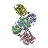


 Mass: 743.405 Da / Num. of mol.: 4 / Source method: isolated from a natural source / Formula: C21H28N7O17P3 / Feature type: SUBJECT OF INVESTIGATION
 Mass: 96.063 Da / Num. of mol.: 8 / Source method: obtained synthetically / Formula: SO4
 Mass: 18.015 Da / Num. of mol.: 232 / Source method: isolated from a natural source / Formula: H2O
 Sample preparation
 / Beamline: I04 / Wavelength: 0.9795 Å
 Processing