 Movie
Movie Controller


+ Open data

- Basic information
| Entry | Database: PDB / ID: 8bjm | |||||||||
|---|---|---|---|---|---|---|---|---|---|---|
| Title | Human full length RAD52 undecamer. | |||||||||
 Components Components | DNA repair protein RAD52 homolog | |||||||||
 Keywords Keywords | DNA BINDING PROTEIN / DNA repair protein / oligomeric structure | |||||||||
| Function / homology |  Function and homology information Function and homology informationdouble-strand break repair via single-strand annealing / DNA double-strand break processing involved in repair via single-strand annealing / DNA recombinase assembly / mitotic recombination / HDR through MMEJ (alt-NHEJ) / regulation of nucleotide-excision repair / HDR through Single Strand Annealing (SSA) / SUMOylation of DNA damage response and repair proteins / protein-DNA complex / double-strand break repair via homologous recombination ...double-strand break repair via single-strand annealing / DNA double-strand break processing involved in repair via single-strand annealing / DNA recombinase assembly / mitotic recombination / HDR through MMEJ (alt-NHEJ) / regulation of nucleotide-excision repair / HDR through Single Strand Annealing (SSA) / SUMOylation of DNA damage response and repair proteins / protein-DNA complex / double-strand break repair via homologous recombination / double-strand break repair / single-stranded DNA binding / cellular response to oxidative stress / DNA recombination / protein-containing complex / DNA binding / nucleoplasm / identical protein binding / nucleus Similarity search - Function | |||||||||
| Biological species |  Homo sapiens (human) Homo sapiens (human) | |||||||||
| Method | ELECTRON MICROSCOPY / single particle reconstruction / cryo EM / Resolution: 2.2 Å | |||||||||
 Authors Authors | Marotta, R. / Balboni, B. / Girotto, S. / Cavalli, A. | |||||||||
| Funding support | European Union,  Italy, 2items Italy, 2items
| |||||||||
 Citation Citation | Journal: Commun Biol / Year: 2024 Title: An integrative structural study of the human full-length RAD52 at 2.2 Å resolution. Authors: Beatrice Balboni / Roberto Marotta / Francesco Rinaldi / Giulia Milordini / Giulia Varignani / Stefania Girotto / Andrea Cavalli /  Abstract: Human RAD52 (RAD52) is a DNA-binding protein involved in many DNA repair mechanisms and genomic stability maintenance. In the last few years, this protein was discovered to be a promising novel ...Human RAD52 (RAD52) is a DNA-binding protein involved in many DNA repair mechanisms and genomic stability maintenance. In the last few years, this protein was discovered to be a promising novel pharmacological target for anticancer strategies. Although the interest in RAD52 has exponentially grown in the previous decade, most information about its structure and mechanism still needs to be elucidated. Here, we report the 2.2 Å resolution cryo-EM reconstruction of the full-length RAD52 (FL-RAD52) protein. This allows us to describe the hydration shell of the N-terminal region of FL-RAD52, which is structured in an undecamer ring. Water molecules coordinate with protein residues to promote stabilization inside and among the protomers and within the inner DNA binding cleft to drive protein-DNA recognition. Additionally, through a multidisciplinary approach involving SEC-SAXS and computational methods, we comprehensively describe the highly flexible and dynamic organization of the C-terminal portion of FL-RAD52. This work discloses unprecedented structural details on the FL-RAD52, which will be critical for characterizing its mechanism of action and inhibitor development, particularly in the context of novel approaches to synthetic lethality and anticancer drug discovery. #1: Journal: Biorxiv / Year: 2023Title: Novel structural insights on full-length human RAD52: Cryo-EM and beyond Authors: Balboni, B. / Marotta, R. / Rinaldi, F. / Girotto, S. / Cavalli, A. | |||||||||
| History |
|
- Structure visualization
| Structure viewer | Molecule: MolmilJmol/JSmol |
|---|
- Downloads & links
-Download
| PDBx/mmCIF format | 8bjm.cif.gz | 439.8 KB | Display | PDBx/mmCIF format |
|---|---|---|---|---|
| PDB format | pdb8bjm.ent.gz | 343.5 KB | Display | PDB format |
| PDBx/mmJSON format | 8bjm.json.gz | Tree view | PDBx/mmJSON format | |
| Others |  Other downloads Other downloads |
-Validation report
| Arichive directory | https://data.pdbj.org/pub/pdb/validation_reports/bj/8bjmftp://data.pdbj.org/pub/pdb/validation_reports/bj/8bjm | HTTPS FTP |
|---|
-Related structure data
| Related structure data |  16089MC M: map data used to model this data C: citing same article ( |
|---|---|
| Similar structure data |
-Links
 PDBj
PDBj


- Assembly
| Deposited unit | 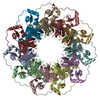
|
|---|---|
| 1 |
|
-Components
| #1: Protein | Mass: 48044.637 Da / Num. of mol.: 11 Source method: isolated from a genetically manipulated source Source: (gene. exp.) Homo sapiens (human) / Gene: RAD52 / Production host:  #2: Water | ChemComp-HOH / |  Mass: 18.015 Da / Num. of mol.: 498 / Source method: isolated from a natural source / Formula: H2O Mass: 18.015 Da / Num. of mol.: 498 / Source method: isolated from a natural source / Formula: H2O |
|---|
-Experimental details
-Experiment
| Experiment | Method: ELECTRON MICROSCOPY |
|---|---|
| EM experiment | Aggregation state: PARTICLE / 3D reconstruction method: single particle reconstruction |
- Sample preparation
| Component | Name: Human full length RAD52 / Type: COMPLEX / Details: N-terminal 6xHis-RAD52 recombinant protein / Entity ID: #1 / Source: RECOMBINANT |
|---|---|
| Molecular weight | Value: 0.650 MDa / Experimental value: YES |
| Source (natural) | Organism: Homo sapiens (human) |
| Source (recombinant) | Organism: |
| Buffer solution | pH: 7.5 |
| Specimen | Conc.: 0.7 mg/ml / Embedding applied: NO / Shadowing applied: NO / Staining applied: NO / Vitrification applied: YES / Details: This sample was monodisperse |
| Specimen support | Grid material: COPPER / Grid mesh size: 300 divisions/in. / Grid type: Quantifoil R1.2/1.3 |
| Vitrification | Instrument: FEI VITROBOT MARK IV / Cryogen name: ETHANE / Humidity: 100 % / Chamber temperature: 318.15 K |
- Electron microscopy imaging
| Experimental equipment |  Model: Titan Krios / Image courtesy: FEI Company |
|---|---|
| Microscopy | Model: FEI TITAN KRIOS |
| Electron gun | Electron source:  FIELD EMISSION GUN / Accelerating voltage: 300 kV / Illumination mode: FLOOD BEAM FIELD EMISSION GUN / Accelerating voltage: 300 kV / Illumination mode: FLOOD BEAM |
| Electron lens | Mode: BRIGHT FIELD / Nominal defocus max: 1800 nm / Nominal defocus min: 800 nm |
| Specimen holder | Cryogen: NITROGEN / Specimen holder model: FEI TITAN KRIOS AUTOGRID HOLDER |
| Image recording | Electron dose: 50 e/Å2 / Film or detector model: FEI FALCON IV (4k x 4k) / Num. of real images: 17400 |
| EM imaging optics | Energyfilter name: TFS Selectris X |
- Processing
| EM software |
| ||||||||||||||||||||||||||||||||||||||||||||
|---|---|---|---|---|---|---|---|---|---|---|---|---|---|---|---|---|---|---|---|---|---|---|---|---|---|---|---|---|---|---|---|---|---|---|---|---|---|---|---|---|---|---|---|---|---|
| CTF correction | Type: PHASE FLIPPING AND AMPLITUDE CORRECTION | ||||||||||||||||||||||||||||||||||||||||||||
| Particle selection | Num. of particles selected: 4124609 | ||||||||||||||||||||||||||||||||||||||||||||
| Symmetry | Point symmetry: C11 (11 fold cyclic) | ||||||||||||||||||||||||||||||||||||||||||||
| 3D reconstruction | Resolution: 2.2 Å / Resolution method: FSC 0.143 CUT-OFF / Num. of particles: 835808 / Num. of class averages: 1 / Symmetry type: POINT | ||||||||||||||||||||||||||||||||||||||||||||
| Refine LS restraints |
|
