 Movie
Movie Controller

[English] 日本語
 Yorodumi
- PDB-8bj2: Crystal structure of Medicago truncatula histidinol-phosphate ami... -
+ Open data

- Basic information
| Entry | Database: PDB / ID: 8bj2 | ||||||
|---|---|---|---|---|---|---|---|
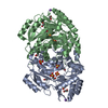
| Title | Crystal structure of Medicago truncatula histidinol-phosphate aminotransferase (HISN6) in the closed state | ||||||
 Components Components | histidinol-phosphate aminotransferase | ||||||
 Keywords Keywords | TRANSFERASE / HISN6 / histidinol-phosphate aminotransferase / aminotransferase / plant / closed | ||||||
| Function / homology |  Function and homology information Function and homology informationhistidinol-phosphate transaminase / histidinol-phosphate transaminase activity / L-histidine biosynthetic process / pyridoxal phosphate binding Similarity search - Function | ||||||
| Biological species |  | ||||||
| Method |  X-RAY DIFFRACTION / SYNCHROTRON / MOLECULAR REPLACEMENT / Resolution: 1.4 Å X-RAY DIFFRACTION / SYNCHROTRON / MOLECULAR REPLACEMENT / Resolution: 1.4 Å | ||||||
 Authors Authors | Rutkiewicz, M. / Ruszkowski, M. | ||||||
| Funding support |  Poland, 1items Poland, 1items
| ||||||
 Citation Citation | Journal: Plant Physiol Biochem. / Year: 2023 Title: Insights into the substrate specificity, structure, and dynamics of plant histidinol-phosphate aminotransferase (HISN6). Authors: Rutkiewicz, M. / Nogues, I. / Witek, W. / Angelaccio, S. / Contestabile, R. / Ruszkowski, M. | ||||||
| History |
|
- Structure visualization
| Structure viewer | Molecule: MolmilJmol/JSmol |
|---|
- Downloads & links
-Download
| PDBx/mmCIF format | 8bj2.cif.gz | 188.6 KB | Display | PDBx/mmCIF format |
|---|---|---|---|---|
| PDB format | pdb8bj2.ent.gz | 145.2 KB | Display | PDB format |
| PDBx/mmJSON format | 8bj2.json.gz | Tree view | PDBx/mmJSON format | |
| Others |  Other downloads Other downloads |
-Validation report
| Summary document | 8bj2_validation.pdf.gz | 894.3 KB | Display | wwPDB validaton report |
|---|---|---|---|---|
| Full document | 8bj2_full_validation.pdf.gz | 897.9 KB | Display | |
| Data in XML | 8bj2_validation.xml.gz | 39 KB | Display | |
| Data in CIF | 8bj2_validation.cif.gz | 61.1 KB | Display | |
| Arichive directory | https://data.pdbj.org/pub/pdb/validation_reports/bj/8bj2ftp://data.pdbj.org/pub/pdb/validation_reports/bj/8bj2 | HTTPS FTP |
-Related structure data


-Links
 PDBj
PDBj- Assembly
| Deposited unit | 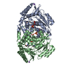
| ||||||||
|---|---|---|---|---|---|---|---|---|---|
| 1 |
| ||||||||
| Unit cell |
|
-Components
| #1: Protein | Mass: 40710.027 Da / Num. of mol.: 2 Source method: isolated from a genetically manipulated source Source: (gene. exp.) Production host:  References: UniProt: A0A072U7F9 #2: Chemical |   Mass: 59.044 Da / Num. of mol.: 2 / Source method: obtained synthetically / Formula: C2H3O2 Mass: 59.044 Da / Num. of mol.: 2 / Source method: obtained synthetically / Formula: C2H3O2#3: Chemical |   Mass: 22.990 Da / Num. of mol.: 2 / Source method: obtained synthetically / Formula: Na Mass: 22.990 Da / Num. of mol.: 2 / Source method: obtained synthetically / Formula: Na#4: Chemical |   Mass: 238.305 Da / Num. of mol.: 2 / Source method: obtained synthetically / Formula: C8H18N2O4S / Feature type: SUBJECT OF INVESTIGATION / Comment: pH buffer*YM Mass: 238.305 Da / Num. of mol.: 2 / Source method: obtained synthetically / Formula: C8H18N2O4S / Feature type: SUBJECT OF INVESTIGATION / Comment: pH buffer*YM#5: Water | ChemComp-HOH / |  Mass: 18.015 Da / Num. of mol.: 1054 / Source method: isolated from a natural source / Formula: H2O Mass: 18.015 Da / Num. of mol.: 1054 / Source method: isolated from a natural source / Formula: H2OHas ligand of interest | Y | |
|---|
-Experimental details
-Experiment
| Experiment | Method: X-RAY DIFFRACTION / Number of used crystals: 1 |
|---|
- Sample preparation
| Crystal | Density Matthews: 2.33 Å3/Da / Density % sol: 47.16 % |
|---|---|
| Crystal grow | Temperature: 292 K / Method: vapor diffusion, hanging drop Details: 0.2M ammonium acetate; 0.1M BIS_TRIS pH 6.5; 25% w/v polyethylene glycol 3350 |
-Data collection
| Diffraction | Mean temperature: 100 K / Serial crystal experiment: N | ||||||||||||||||||||||||||||||||||||||||||||||||||||||||||||||||||||||||||||||||||||||||||||||||||||
|---|---|---|---|---|---|---|---|---|---|---|---|---|---|---|---|---|---|---|---|---|---|---|---|---|---|---|---|---|---|---|---|---|---|---|---|---|---|---|---|---|---|---|---|---|---|---|---|---|---|---|---|---|---|---|---|---|---|---|---|---|---|---|---|---|---|---|---|---|---|---|---|---|---|---|---|---|---|---|---|---|---|---|---|---|---|---|---|---|---|---|---|---|---|---|---|---|---|---|---|---|---|
| Diffraction source | Source: SYNCHROTRON / Site: APS  / Beamline: 22-ID / Wavelength: 1 Å / Beamline: 22-ID / Wavelength: 1 Å | ||||||||||||||||||||||||||||||||||||||||||||||||||||||||||||||||||||||||||||||||||||||||||||||||||||
| Detector | Type: DECTRIS EIGER X 16M / Detector: PIXEL / Date: Dec 18, 2018 | ||||||||||||||||||||||||||||||||||||||||||||||||||||||||||||||||||||||||||||||||||||||||||||||||||||
| Radiation | Protocol: SINGLE WAVELENGTH / Monochromatic (M) / Laue (L): M / Scattering type: x-ray | ||||||||||||||||||||||||||||||||||||||||||||||||||||||||||||||||||||||||||||||||||||||||||||||||||||
| Radiation wavelength | Wavelength: 1 Å / Relative weight: 1 | ||||||||||||||||||||||||||||||||||||||||||||||||||||||||||||||||||||||||||||||||||||||||||||||||||||
| Reflection | Resolution: 1.4→53.86 Å / Num. obs: 145641 / % possible obs: 99.2 % / Redundancy: 4.027 % / Biso Wilson estimate: 16.77 Å2 / CC1/2: 0.999 / Rmerge(I) obs: 0.05 / Rrim(I) all: 0.058 / Χ2: 1.044 / Net I/σ(I): 13.89 / Num. measured all: 586491 | ||||||||||||||||||||||||||||||||||||||||||||||||||||||||||||||||||||||||||||||||||||||||||||||||||||
| Reflection shell | Diffraction-ID: 1
|
- Processing
| Software |
| |||||||||||||||||||||||||||||||||||||||||||||||||||||||||||||||
|---|---|---|---|---|---|---|---|---|---|---|---|---|---|---|---|---|---|---|---|---|---|---|---|---|---|---|---|---|---|---|---|---|---|---|---|---|---|---|---|---|---|---|---|---|---|---|---|---|---|---|---|---|---|---|---|---|---|---|---|---|---|---|---|---|
| Refinement | Method to determine structure: MOLECULAR REPLACEMENT Starting model: MtHISN6_open Resolution: 1.4→53.86 Å / SU ML: 0.14 / Cross valid method: FREE R-VALUE / σ(F): 1.36 / Phase error: 17.23 / Stereochemistry target values: ML
| |||||||||||||||||||||||||||||||||||||||||||||||||||||||||||||||
| Solvent computation | Shrinkage radii: 0.9 Å / VDW probe radii: 1.11 Å / Solvent model: FLAT BULK SOLVENT MODEL | |||||||||||||||||||||||||||||||||||||||||||||||||||||||||||||||
| Displacement parameters | Biso max: 54.57 Å2 / Biso mean: 21.2318 Å2 / Biso min: 11.77 Å2 | |||||||||||||||||||||||||||||||||||||||||||||||||||||||||||||||
| Refinement step | Cycle: final / Resolution: 1.4→53.86 Å
| |||||||||||||||||||||||||||||||||||||||||||||||||||||||||||||||
| LS refinement shell | Refine-ID: X-RAY DIFFRACTION / Rfactor Rfree error: 0 / Total num. of bins used: 8
|
