[English] 日本語
 Yorodumi
- PDB-8bgw: CryoEM structure of quinol-dependent Nitric Oxide Reductase (qNOR... -
+ Open data
- Basic information
| Entry | Database: PDB / ID: 8bgw | |||||||||
|---|---|---|---|---|---|---|---|---|---|---|
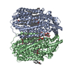
| Title | CryoEM structure of quinol-dependent Nitric Oxide Reductase (qNOR) from Alcaligenes xylosoxidans at 2.2 A resolution | |||||||||
 Components Components | Nitric oxide reductase subunit B | |||||||||
 Keywords Keywords | MEMBRANE PROTEIN / quinol-dependent Nitric Oxide Reductase / proton transfer / quinol binding / ubiquinol oxidase | |||||||||
| Function / homology |  Function and homology information Function and homology informationnitric oxide reductase (cytochrome c) / nitric oxide reductase activity / cytochrome-c oxidase activity / aerobic respiration / heme binding / membrane Similarity search - Function | |||||||||
| Biological species |  Achromobacter xylosoxidans (bacteria) Achromobacter xylosoxidans (bacteria) | |||||||||
| Method | ELECTRON MICROSCOPY / single particle reconstruction / cryo EM / Resolution: 2.2 Å | |||||||||
 Authors Authors | Flynn, A. / Antonyuk, S.V. / Eady, R.R. / Muench, S.P. / Hasnain, S.S. | |||||||||
| Funding support |  United Kingdom, 2items United Kingdom, 2items
| |||||||||
 Citation Citation |  Journal: Nat Commun / Year: 2023 Journal: Nat Commun / Year: 2023Title: A 2.2 Å cryoEM structure of a quinol-dependent NO Reductase shows close similarity to respiratory oxidases. Authors: Alex J Flynn / Svetlana V Antonyuk / Robert R Eady / Stephen P Muench / S Samar Hasnain /  Abstract: Quinol-dependent nitric oxide reductases (qNORs) are considered members of the respiratory heme-copper oxidase superfamily, are unique to bacteria, and are commonly found in pathogenic bacteria where ...Quinol-dependent nitric oxide reductases (qNORs) are considered members of the respiratory heme-copper oxidase superfamily, are unique to bacteria, and are commonly found in pathogenic bacteria where they play a role in combating the host immune response. qNORs are also essential enzymes in the denitrification pathway, catalysing the reduction of nitric oxide to nitrous oxide. Here, we determine a 2.2 Å cryoEM structure of qNOR from Alcaligenes xylosoxidans, an opportunistic pathogen and a denitrifying bacterium of importance in the nitrogen cycle. This high-resolution structure provides insight into electron, substrate, and proton pathways, and provides evidence that the quinol binding site not only contains the conserved His and Asp residues but also possesses a critical Arg (Arg720) observed in cytochrome bo, a respiratory quinol oxidase. | |||||||||
| History |
|
- Structure visualization
| Structure viewer | Molecule:  Molmil Molmil Jmol/JSmol Jmol/JSmol |
|---|
- Downloads & links
- Download
| PDBx/mmCIF format |  8bgw.cif.gz 8bgw.cif.gz | 332 KB | Display |  PDBx/mmCIF format PDBx/mmCIF format |
|---|---|---|---|---|
| PDB format |  pdb8bgw.ent.gz pdb8bgw.ent.gz | 265.9 KB | Display |  PDB format PDB format |
| PDBx/mmJSON format |  8bgw.json.gz 8bgw.json.gz | Tree view |  PDBx/mmJSON format PDBx/mmJSON format | |
| Others |  Other downloads Other downloads |
-Validation report
| Summary document |  8bgw_validation.pdf.gz 8bgw_validation.pdf.gz | 1.8 MB | Display |  wwPDB validaton report wwPDB validaton report |
|---|---|---|---|---|
| Full document |  8bgw_full_validation.pdf.gz 8bgw_full_validation.pdf.gz | 1.9 MB | Display | |
| Data in XML |  8bgw_validation.xml.gz 8bgw_validation.xml.gz | 69.8 KB | Display | |
| Data in CIF |  8bgw_validation.cif.gz 8bgw_validation.cif.gz | 101.7 KB | Display | |
| Arichive directory |  https://data.pdbj.org/pub/pdb/validation_reports/bg/8bgw https://data.pdbj.org/pub/pdb/validation_reports/bg/8bgw ftp://data.pdbj.org/pub/pdb/validation_reports/bg/8bgw ftp://data.pdbj.org/pub/pdb/validation_reports/bg/8bgw | HTTPS FTP |
-Related structure data
| Related structure data |  16041MC M: map data used to model this data C: citing same article ( |
|---|---|
| Similar structure data | Similarity search - Function & homology  F&H Search F&H Search |
- Links
Links
- Assembly
| Deposited unit | 
| |||||||||
|---|---|---|---|---|---|---|---|---|---|---|
| 1 |
| |||||||||
| Noncrystallographic symmetry (NCS) | NCS domain:
NCS ensembles : (Details: Local NCS retraints between domains: 1 2) |
- Components
-Protein / Sugars , 2 types, 4 molecules AB

| #1: Protein | Mass: 84724.867 Da / Num. of mol.: 2 Source method: isolated from a genetically manipulated source Source: (gene. exp.)  Achromobacter xylosoxidans (bacteria) / Gene: norB_1, ERS451415_02175, I6G72_25885 / Plasmid: PET-26B (+) / Production host: Achromobacter xylosoxidans (bacteria) / Gene: norB_1, ERS451415_02175, I6G72_25885 / Plasmid: PET-26B (+) / Production host:  References: UniProt: A0A0D6H8R3, nitric oxide reductase (cytochrome c) #5: Sugar | |
|---|
-Non-polymers , 6 types, 472 molecules 


| #2: Chemical | ChemComp-HEM / #3: Chemical | #4: Chemical | #6: Chemical | #7: Chemical | ChemComp-LOP / ( #8: Water | ChemComp-HOH / | |
|---|
-Details
| Has ligand of interest | Y |
|---|
-Experimental details
-Experiment
| Experiment | Method: ELECTRON MICROSCOPY |
|---|---|
| EM experiment | Aggregation state: PARTICLE / 3D reconstruction method: single particle reconstruction |
- Sample preparation
| Component | Name: quinol-dependent Nitric Oxide Reductase / Type: CELL / Details: quinol-dependent Nitric Oxide Reductase / Entity ID: #1 / Source: RECOMBINANT |
|---|---|
| Molecular weight | Value: 0.84651 MDa / Experimental value: NO |
| Source (natural) | Organism:  Achromobacter xylosoxidans (bacteria) Achromobacter xylosoxidans (bacteria) |
| Source (recombinant) | Organism:  |
| Buffer solution | pH: 7 / Details: 50mM Tris-HCl, 150mM NaCl, 0.05% DTM |
| Buffer component | Conc.: 50 mM / Name: Tris-HCL / Formula: Tris-HCL |
| Specimen | Conc.: 3 mg/ml / Embedding applied: NO / Shadowing applied: NO / Staining applied: NO / Vitrification applied: YES / Details: Sample was monodisperse |
| Specimen support | Grid material: GOLD / Grid mesh size: 300 divisions/in. / Grid type: Quantifoil R1.2/1.3 |
| Vitrification | Instrument: FEI VITROBOT MARK IV / Cryogen name: ETHANE / Humidity: 100 % / Chamber temperature: 277 K Details: Sample at 3 mg/mL was applied to glow-discharged Quantifoil Au R1.2/1.3 holey carbo grids |
- Electron microscopy imaging
| Experimental equipment |  Model: Titan Krios / Image courtesy: FEI Company |
|---|---|
| Microscopy | Model: FEI TITAN KRIOS |
| Electron gun | Electron source:  FIELD EMISSION GUN / Accelerating voltage: 300 kV / Illumination mode: SPOT SCAN FIELD EMISSION GUN / Accelerating voltage: 300 kV / Illumination mode: SPOT SCAN |
| Electron lens | Mode: BRIGHT FIELD / Nominal magnification: 130000 X / Nominal defocus max: 2700 nm / Nominal defocus min: 900 nm / Cs: 2.7 mm / C2 aperture diameter: 50 µm / Alignment procedure: COMA FREE |
| Specimen holder | Cryogen: NITROGEN / Specimen holder model: FEI TITAN KRIOS AUTOGRID HOLDER / Temperature (max): 114 K / Temperature (min): 80 K |
| Image recording | Average exposure time: 6.11 sec. / Electron dose: 34.9 e/Å2 / Film or detector model: FEI FALCON IV (4k x 4k) / Num. of grids imaged: 1 / Num. of real images: 5466 |
| EM imaging optics | Energyfilter name: TFS Selectris / Energyfilter slit width: 10 eV |
| Image scans | Width: 4096 / Height: 4096 |
- Processing
| Software | Name: REFMAC / Version: 5.8.0267 / Classification: refinement | ||||||||||||||||||||||||||||||||||||||||||||||||||||||||||||||||||||||||||||||||||||||||||||||||||||||||||
|---|---|---|---|---|---|---|---|---|---|---|---|---|---|---|---|---|---|---|---|---|---|---|---|---|---|---|---|---|---|---|---|---|---|---|---|---|---|---|---|---|---|---|---|---|---|---|---|---|---|---|---|---|---|---|---|---|---|---|---|---|---|---|---|---|---|---|---|---|---|---|---|---|---|---|---|---|---|---|---|---|---|---|---|---|---|---|---|---|---|---|---|---|---|---|---|---|---|---|---|---|---|---|---|---|---|---|---|
| EM software |
| ||||||||||||||||||||||||||||||||||||||||||||||||||||||||||||||||||||||||||||||||||||||||||||||||||||||||||
| CTF correction | Type: PHASE FLIPPING AND AMPLITUDE CORRECTION | ||||||||||||||||||||||||||||||||||||||||||||||||||||||||||||||||||||||||||||||||||||||||||||||||||||||||||
| Particle selection | Num. of particles selected: 3000000 | ||||||||||||||||||||||||||||||||||||||||||||||||||||||||||||||||||||||||||||||||||||||||||||||||||||||||||
| Symmetry | Point symmetry: C2 (2 fold cyclic) | ||||||||||||||||||||||||||||||||||||||||||||||||||||||||||||||||||||||||||||||||||||||||||||||||||||||||||
| 3D reconstruction | Resolution: 2.2 Å / Resolution method: FSC 0.143 CUT-OFF / Num. of particles: 404950 / Algorithm: FOURIER SPACE / Num. of class averages: 2 / Symmetry type: POINT | ||||||||||||||||||||||||||||||||||||||||||||||||||||||||||||||||||||||||||||||||||||||||||||||||||||||||||
| Atomic model building | B value: 47 / Protocol: RIGID BODY FIT / Space: RECIPROCAL / Target criteria: Cross-correlation coefficient | ||||||||||||||||||||||||||||||||||||||||||||||||||||||||||||||||||||||||||||||||||||||||||||||||||||||||||
| Refinement | Highest resolution: 2.2 Å | ||||||||||||||||||||||||||||||||||||||||||||||||||||||||||||||||||||||||||||||||||||||||||||||||||||||||||
| Refine LS restraints |
|
 Movie
Movie Controller


 PDBj
PDBj


