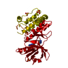
- PDB-8bfm: Crystal structure of human calmodulin-dependent protein kinase 1D... -
+
Open data
ID or keywords:
Loading...
-
Basic information
Entry
Database: PDB / ID: 8bfm
Title
Crystal structure of human calmodulin-dependent protein kinase 1D (CAMK1D) in complex with FZ331
Components
Calcium/calmodulin-dependent protein kinase type 1D
Keywords
TRANSFERASE / CAMK1D / Kinase
Function / homology
Function and homology information
regulation of granulocyte chemotaxis / : / regulation of dendrite development / Ca2+/calmodulin-dependent protein kinase / calcium/calmodulin-dependent protein kinase activity / positive regulation of neutrophil chemotaxis / regulation of neuron projection development / positive regulation of respiratory burst / positive regulation of phagocytosis / positive regulation of neuron projection development ...regulation of granulocyte chemotaxis / : / regulation of dendrite development / Ca2+/calmodulin-dependent protein kinase / calcium/calmodulin-dependent protein kinase activity / positive regulation of neutrophil chemotaxis / regulation of neuron projection development / positive regulation of respiratory burst / positive regulation of phagocytosis / positive regulation of neuron projection development / nervous system development / calmodulin binding / positive regulation of apoptotic process / inflammatory response / protein serine kinase activity / negative regulation of apoptotic process / signal transduction / ATP binding / nucleus / cytoplasm Similarity search - Function
Serine/threonine-protein kinase, active site / Serine/Threonine protein kinases active-site signature. / Protein kinase domain / Serine/Threonine protein kinases, catalytic domain / Protein kinase, ATP binding site / Protein kinases ATP-binding region signature. / Protein kinase domain profile. / Protein kinase domain / Protein kinase-like domain superfamily Similarity search - Domain/homology
pyrazolo[5,1-a]phthalazin-6-amine / Calcium/calmodulin-dependent protein kinase type 1D Similarity search - Component
Calcium/calmodulin-dependentproteinkinasetype1D / CaM kinase I delta / CaM kinase ID / CaM-KI delta / CaMKI delta / CaMKID / CaMKI-like protein kinase / CKLiK
Mass: 42972.801 Da / Num. of mol.: 1 Source method: isolated from a genetically manipulated source Source: (gene. exp.) Homo sapiens (human) / Gene: CAMK1D, CAMKID / Production host: Escherichia coli (E. coli) References: UniProt: Q8IU85, Ca2+/calmodulin-dependent protein kinase
Resolution: 1.7→44.95 Å / Cor.coef. Fo:Fc: 0.963 / Cor.coef. Fo:Fc free: 0.956 / SU B: 5.937 / SU ML: 0.093 / SU R Cruickshank DPI: 0.1223 / Cross valid method: THROUGHOUT / σ(F): 0 / ESU R: 0.122 / ESU R Free: 0.112 / Stereochemistry target values: MAXIMUM LIKELIHOOD Details: HYDROGENS HAVE BEEN ADDED IN THE RIDING POSITIONS U VALUES : WITH TLS ADDED
Rfactor
Num. reflection
% reflection
Selection details
Rfree
0.2242
1427
5 %
RANDOM
Rwork
0.2
-
-
-
obs
0.2012
27070
95.85 %
-
Solvent computation
Ion probe radii: 0.8 Å / Shrinkage radii: 0.8 Å / VDW probe radii: 1.2 Å / Solvent model: MASK
In the structure databanks used in Yorodumi, some data are registered as the other names, "COVID-19 virus" and "2019-nCoV". Here are the details of the virus and the list of structure data.
Jan 31, 2019. EMDB accession codes are about to change! (news from PDBe EMDB page)
EMDB accession codes are about to change! (news from PDBe EMDB page)
The allocation of 4 digits for EMDB accession codes will soon come to an end. Whilst these codes will remain in use, new EMDB accession codes will include an additional digit and will expand incrementally as the available range of codes is exhausted. The current 4-digit format prefixed with “EMD-” (i.e. EMD-XXXX) will advance to a 5-digit format (i.e. EMD-XXXXX), and so on. It is currently estimated that the 4-digit codes will be depleted around Spring 2019, at which point the 5-digit format will come into force.
The EM Navigator/Yorodumi systems omit the EMD- prefix.
Related info.:Q: What is EMD? / ID/Accession-code notation in Yorodumi/EM Navigator
Yorodumi is a browser for structure data from EMDB, PDB, SASBDB, etc.
This page is also the successor to EM Navigator detail page, and also detail information page/front-end page for Omokage search.
The word "yorodu" (or yorozu) is an old Japanese word meaning "ten thousand". "mi" (miru) is to see.
Related info.:EMDB / PDB / SASBDB / Comparison of 3 databanks / Yorodumi Search / Aug 31, 2016. New EM Navigator & Yorodumi / Yorodumi Papers / Jmol/JSmol / Function and homology information / Changes in new EM Navigator and Yorodumi
 Movie
Movie Controller

 Yorodumi
 Open data
 Basic information
 Components
 Keywords
 Function and homology information
 Homo sapiens (human)
 X-RAY DIFFRACTION /
  Authors
 Canada, 1items
  Citation
 Structure visualization
 Downloads & links
 Other downloads

 PDBj
PDBj
 Assembly
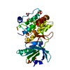


 Mass: 96.063 Da / Num. of mol.: 5 / Source method: obtained synthetically / Formula: SO4
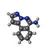
 Mass: 184.197 Da / Num. of mol.: 2 / Source method: obtained synthetically / Formula: C10H8N4 / Feature type: SUBJECT OF INVESTIGATION
 Mass: 18.015 Da / Num. of mol.: 113 / Source method: isolated from a natural source / Formula: H2O
 Sample preparation
 / Beamline: X06SA / Wavelength: 0.99999 Å
 Processing