Netherlands Organisation for Scientific Research (NWO)
84.034.014
Netherlands
Citation
Journal: Nat Commun / Year: 2022 Title: High-resolution reconstruction of a Jumbo-bacteriophage infecting capsulated bacteria using hyperbranched tail fibers. Authors: Ruochen Ouyang / Ana Rita Costa / C Keith Cassidy / Aleksandra Otwinowska / Vera C J Williams / Agnieszka Latka / Phill J Stansfeld / Zuzanna Drulis-Kawa / Yves Briers / Daniël M Pelt / ...Authors: Ruochen Ouyang / Ana Rita Costa / C Keith Cassidy / Aleksandra Otwinowska / Vera C J Williams / Agnieszka Latka / Phill J Stansfeld / Zuzanna Drulis-Kawa / Yves Briers / Daniël M Pelt / Stan J J Brouns / Ariane Briegel / Abstract: The Klebsiella jumbo myophage ϕKp24 displays an unusually complex arrangement of tail fibers interacting with a host cell. In this study, we combine cryo-electron microscopy methods, protein ...The Klebsiella jumbo myophage ϕKp24 displays an unusually complex arrangement of tail fibers interacting with a host cell. In this study, we combine cryo-electron microscopy methods, protein structure prediction methods, molecular simulations, microbiological and machine learning approaches to explore the capsid, tail, and tail fibers of ϕKp24. We determine the structure of the capsid and tail at 4.1 Å and 3.0 Å resolution. We observe the tail fibers are branched and rearranged dramatically upon cell surface attachment. This complex configuration involves fourteen putative tail fibers with depolymerase activity that provide ϕKp24 with the ability to infect a broad panel of capsular polysaccharide (CPS) types of Klebsiella pneumoniae. Our study provides structural and functional insight into how ϕKp24 adapts to the variable surfaces of capsulated bacterial pathogens, which is useful for the development of phage therapy approaches against pan-drug resistant K. pneumoniae strains.
History
Deposition
Oct 26, 2022
Deposition site: PDBE / Processing site: PDBE
Revision 1.0
Dec 7, 2022
Provider: repository / Type: Initial release
Revision 1.1
Jul 24, 2024
Group: Data collection / Category: chem_comp_atom / chem_comp_bond
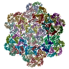
A: Putative virion structural protein B: Putative virion structural protein C: Putative virion structural protein D: Putative virion structural protein E: Putative virion structural protein F: Putative virion structural protein G: Putative virion structural protein H: Putative virion structural protein I: Putative virion structural protein J: Putative virion structural protein K: Putative virion structural protein L: Putative virion structural protein M: Putative virion structural protein N: Putative virion structural protein O: Putative virion structural protein P: Putative virion structural protein Q: Putative virion structural protein R: Putative virion structural protein
In the structure databanks used in Yorodumi, some data are registered as the other names, "COVID-19 virus" and "2019-nCoV". Here are the details of the virus and the list of structure data.
Jan 31, 2019. EMDB accession codes are about to change! (news from PDBe EMDB page)
EMDB accession codes are about to change! (news from PDBe EMDB page)
The allocation of 4 digits for EMDB accession codes will soon come to an end. Whilst these codes will remain in use, new EMDB accession codes will include an additional digit and will expand incrementally as the available range of codes is exhausted. The current 4-digit format prefixed with “EMD-” (i.e. EMD-XXXX) will advance to a 5-digit format (i.e. EMD-XXXXX), and so on. It is currently estimated that the 4-digit codes will be depleted around Spring 2019, at which point the 5-digit format will come into force.
The EM Navigator/Yorodumi systems omit the EMD- prefix.
Related info.:Q: What is EMD? / ID/Accession-code notation in Yorodumi/EM Navigator
Yorodumi is a browser for structure data from EMDB, PDB, SASBDB, etc.
This page is also the successor to EM Navigator detail page, and also detail information page/front-end page for Omokage search.
The word "yorodu" (or yorozu) is an old Japanese word meaning "ten thousand". "mi" (miru) is to see.
Related info.:EMDB / PDB / SASBDB / Comparison of 3 databanks / Yorodumi Search / Aug 31, 2016. New EM Navigator & Yorodumi / Yorodumi Papers / Jmol/JSmol / Function and homology information / Changes in new EM Navigator and Yorodumi
 Movie
Movie Controller


 Open data
 Basic information
 Components
 Keywords
 Function and homology information
 Klebsiella phage vB_KpM_FBKp24 (virus)
 Authors
 Netherlands, 1items
  Citation


 Structure visualization
 Downloads & links
 Other downloads


 PDBj
PDBj Assembly
 Sample preparation
 Electron microscopy imaging
 Processing