 Movie
Movie Controller

[English] 日本語
 Yorodumi
- PDB-8bes: Structure of D188A-fructofuranosidase from Rhodotorula dairenensi... -
+ Open data

- Basic information
| Entry | Database: PDB / ID: 8bes | ||||||
|---|---|---|---|---|---|---|---|
| Title | Structure of D188A-fructofuranosidase from Rhodotorula dairenensis in complex with fructose | ||||||
 Components Components | Beta-fructofuranosidase | ||||||
 Keywords Keywords | HYDROLASE / FRUCTOFURANOSIDASE / INVERTASE / HYDROLYTIC ENZYME / GLYCOSIL HYDROLASE / PREBIOTIC / OLIGOSACCHARIDE / FRUCTOOLIGOSACCHARIDE / FOS / RHODOTORULA DAIRENENSIS | ||||||
| Function / homology |  Function and homology information Function and homology informationbeta-fructofuranosidase / sucrose catabolic process / sucrose alpha-glucosidase activity / fungal-type vacuole Similarity search - Function | ||||||
| Biological species |  Rhodotorula dairenensis (fungus) Rhodotorula dairenensis (fungus) | ||||||
| Method |  X-RAY DIFFRACTION / SYNCHROTRON / FOURIER SYNTHESIS / Resolution: 1.86 Å X-RAY DIFFRACTION / SYNCHROTRON / FOURIER SYNTHESIS / Resolution: 1.86 Å | ||||||
 Authors Authors | Jimenez-Ortega, E. / Sanz-Aparicio, J. | ||||||
| Funding support |  Spain, 1items Spain, 1items
| ||||||
 Citation Citation | Journal: Int J Mol Sci / Year: 2022 Title: Insights into the Structure of the Highly Glycosylated Ffase from Rhodotorula dairenensis Enhance Its Biotechnological Potential. Authors: Jimenez-Ortega, E. / Narmontaite, E. / Gonzalez-Perez, B. / Plou, F.J. / Fernandez-Lobato, M. / Sanz-Aparicio, J. | ||||||
| History |
|
- Structure visualization
| Structure viewer | Molecule: MolmilJmol/JSmol |
|---|
- Downloads & links
-Download
| PDBx/mmCIF format | 8bes.cif.gz | 482.5 KB | Display | PDBx/mmCIF format |
|---|---|---|---|---|
| PDB format | pdb8bes.ent.gz | 390.3 KB | Display | PDB format |
| PDBx/mmJSON format | 8bes.json.gz | Tree view | PDBx/mmJSON format | |
| Others |  Other downloads Other downloads |
-Validation report
| Arichive directory | https://data.pdbj.org/pub/pdb/validation_reports/be/8besftp://data.pdbj.org/pub/pdb/validation_reports/be/8bes | HTTPS FTP |
|---|
-Related structure data
| Related structure data | 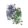 8beqSC  8betC  8beuC S: Starting model for refinement C: citing same article ( |
|---|---|
| Similar structure data |
-Links
 PDBj
PDBj


- Assembly
| Deposited unit | 
| ||||||||
|---|---|---|---|---|---|---|---|---|---|
| 1 | 
| ||||||||
| 2 | 
| ||||||||
| Unit cell |
|
-Components
-Protein , 1 types, 4 molecules ABCD
| #1: Protein | Mass: 70671.266 Da / Num. of mol.: 4 Source method: isolated from a genetically manipulated source Source: (gene. exp.) Rhodotorula dairenensis (fungus) / Gene: INV / Production host: Komagataella pastoris (fungus) / Strain (production host): GS115 / References: UniProt: A0A856TAI5, beta-fructofuranosidase |
|---|
-Sugars , 7 types, 53 molecules 

| #2: Polysaccharide | alpha-D-mannopyranose-(1-3)-beta-D-mannopyranose-(1-4)-2-acetamido-2-deoxy-beta-D-glucopyranose-(1- ...alpha-D-mannopyranose-(1-3)-beta-D-mannopyranose-(1-4)-2-acetamido-2-deoxy-beta-D-glucopyranose-(1-4)-2-acetamido-2-deoxy-beta-D-glucopyranose Source method: isolated from a genetically manipulated source | ||||||||||
|---|---|---|---|---|---|---|---|---|---|---|---|
| #3: Polysaccharide | Source method: isolated from a genetically manipulated source #4: Polysaccharide | 2-acetamido-2-deoxy-beta-D-glucopyranose-(1-4)-2-acetamido-2-deoxy-beta-D-glucopyranose Source method: isolated from a genetically manipulated source #5: Polysaccharide | alpha-D-mannopyranose-(1-3)-alpha-D-mannopyranose-(1-6)-[alpha-D-mannopyranose-(1-3)]beta-D- ...alpha-D-mannopyranose-(1-3)-alpha-D-mannopyranose-(1-6)-[alpha-D-mannopyranose-(1-3)]beta-D-mannopyranose-(1-4)-2-acetamido-2-deoxy-beta-D-glucopyranose-(1-4)-2-acetamido-2-deoxy-beta-D-glucopyranose | Source method: isolated from a genetically manipulated source #6: Sugar | ChemComp-MAN /  Type: D-saccharide, alpha linking / Mass: 180.156 Da / Num. of mol.: 13 / Source method: obtained synthetically / Formula: C6H12O6 Type: D-saccharide, alpha linking / Mass: 180.156 Da / Num. of mol.: 13 / Source method: obtained synthetically / Formula: C6H12O6#7: Sugar | ChemComp-NAG /  Type: D-saccharide, beta linking / Mass: 221.208 Da / Num. of mol.: 28 / Source method: obtained synthetically / Formula: C8H15NO6 Type: D-saccharide, beta linking / Mass: 221.208 Da / Num. of mol.: 28 / Source method: obtained synthetically / Formula: C8H15NO6#9: Sugar | ChemComp-FRU /  Type: D-saccharide, beta linking / Mass: 180.156 Da / Num. of mol.: 4 / Source method: obtained synthetically / Formula: C6H12O6 / Feature type: SUBJECT OF INVESTIGATION Type: D-saccharide, beta linking / Mass: 180.156 Da / Num. of mol.: 4 / Source method: obtained synthetically / Formula: C6H12O6 / Feature type: SUBJECT OF INVESTIGATION |
-Non-polymers , 3 types, 1977 molecules 


| #8: Chemical | ChemComp-BTB /  Mass: 209.240 Da / Num. of mol.: 4 / Source method: obtained synthetically / Formula: C8H19NO5 / Comment: pH buffer*YM Mass: 209.240 Da / Num. of mol.: 4 / Source method: obtained synthetically / Formula: C8H19NO5 / Comment: pH buffer*YM#10: Chemical | ChemComp-PEG / |  Mass: 106.120 Da / Num. of mol.: 1 / Source method: obtained synthetically / Formula: C4H10O3 Mass: 106.120 Da / Num. of mol.: 1 / Source method: obtained synthetically / Formula: C4H10O3#11: Water | ChemComp-HOH / | Mass: 18.015 Da / Num. of mol.: 1972 / Source method: isolated from a natural source / Formula: H2O |
|---|
-Details
| Has ligand of interest | Y |
|---|---|
| Has protein modification | Y |
-Experimental details
-Experiment
| Experiment | Method: X-RAY DIFFRACTION / Number of used crystals: 1 |
|---|
- Sample preparation
| Crystal | Density Matthews: 2.54 Å3/Da / Density % sol: 51.67 % / Description: Blade crystal |
|---|---|
| Crystal grow | Temperature: 291 K / Method: vapor diffusion, sitting drop / pH: 5.5 Details: 18% PEG 3350, 0.2 M MgCl2, 0.1M Bis-Tris pH5.5 at 5.1 mg ml-1 concentration in presence of 20 mM fructose, 1:1 rate Cryocooling: Mother liquor solution supplemented with 20 mM fructose and 20% glycerol |
-Data collection
| Diffraction | Mean temperature: 100 K / Serial crystal experiment: N |
|---|---|
| Diffraction source | Source: SYNCHROTRON / Site: ALBA / Beamline: XALOC / Wavelength: 0.979257 Å |
| Detector | Type: DECTRIS PILATUS 6M / Detector: PIXEL / Date: Jul 22, 2021 / Details: KB Mirrors |
| Radiation | Monochromator: Si(111) channel-cut, cryocooler / Protocol: SINGLE WAVELENGTH / Monochromatic (M) / Laue (L): M / Scattering type: x-ray |
| Radiation wavelength | Wavelength: 0.979257 Å / Relative weight: 1 |
| Reflection | Resolution: 1.86→45.74 Å / Num. obs: 202338 / % possible obs: 99.9 % / Redundancy: 5.5 % / CC1/2: 0.998 / Rmerge(I) obs: 0.071 / Rpim(I) all: 0.033 / Rrim(I) all: 0.078 / Net I/σ(I): 14 |
| Reflection shell | Resolution: 1.86→1.89 Å / Redundancy: 5.7 % / Rmerge(I) obs: 0.612 / Mean I/σ(I) obs: 2.8 / Num. unique obs: 10039 / CC1/2: 0.833 / Rpim(I) all: 0.399 / % possible all: 100 |
- Processing
| Software |
| ||||||||||||||||||||||||||||||||||||||||||||||||||||||||||||||||||||||||||||||||||||||||||||||||||||||||||||||||||||||||||||||||||||||||||||||||||||||||||||||||||||||||||||||||||||||
|---|---|---|---|---|---|---|---|---|---|---|---|---|---|---|---|---|---|---|---|---|---|---|---|---|---|---|---|---|---|---|---|---|---|---|---|---|---|---|---|---|---|---|---|---|---|---|---|---|---|---|---|---|---|---|---|---|---|---|---|---|---|---|---|---|---|---|---|---|---|---|---|---|---|---|---|---|---|---|---|---|---|---|---|---|---|---|---|---|---|---|---|---|---|---|---|---|---|---|---|---|---|---|---|---|---|---|---|---|---|---|---|---|---|---|---|---|---|---|---|---|---|---|---|---|---|---|---|---|---|---|---|---|---|---|---|---|---|---|---|---|---|---|---|---|---|---|---|---|---|---|---|---|---|---|---|---|---|---|---|---|---|---|---|---|---|---|---|---|---|---|---|---|---|---|---|---|---|---|---|---|---|---|---|
| Refinement | Method to determine structure: FOURIER SYNTHESIS Starting model: 8BEQ Resolution: 1.86→45.74 Å / Cor.coef. Fo:Fc: 0.961 / Cor.coef. Fo:Fc free: 0.945 / SU B: 3.193 / SU ML: 0.094 / Cross valid method: THROUGHOUT / ESU R: 0.142 / ESU R Free: 0.13 / Stereochemistry target values: MAXIMUM LIKELIHOOD / Details: HYDROGENS HAVE BEEN ADDED IN THE RIDING POSITIONS
| ||||||||||||||||||||||||||||||||||||||||||||||||||||||||||||||||||||||||||||||||||||||||||||||||||||||||||||||||||||||||||||||||||||||||||||||||||||||||||||||||||||||||||||||||||||||
| Solvent computation | Ion probe radii: 0.8 Å / Shrinkage radii: 0.8 Å / VDW probe radii: 1.2 Å / Solvent model: MASK | ||||||||||||||||||||||||||||||||||||||||||||||||||||||||||||||||||||||||||||||||||||||||||||||||||||||||||||||||||||||||||||||||||||||||||||||||||||||||||||||||||||||||||||||||||||||
| Displacement parameters | Biso mean: 26.497 Å2
| ||||||||||||||||||||||||||||||||||||||||||||||||||||||||||||||||||||||||||||||||||||||||||||||||||||||||||||||||||||||||||||||||||||||||||||||||||||||||||||||||||||||||||||||||||||||
| Refinement step | Cycle: 1 / Resolution: 1.86→45.74 Å
| ||||||||||||||||||||||||||||||||||||||||||||||||||||||||||||||||||||||||||||||||||||||||||||||||||||||||||||||||||||||||||||||||||||||||||||||||||||||||||||||||||||||||||||||||||||||
| Refine LS restraints |
|