 Movie
Movie Controller

[English] 日本語
 Yorodumi
- PDB-8bbr: Determination of the structure of active tyrosinase from bacteriu... -
+ Open data

- Basic information
| Entry | Database: PDB / ID: 8bbr | ||||||
|---|---|---|---|---|---|---|---|
| Title | Determination of the structure of active tyrosinase from bacterium Verrucomicrobium spinosum | ||||||
 Components Components | Core tyrosinase | ||||||
 Keywords Keywords | METAL BINDING PROTEIN / Tyrosinase / Copper / Verrucomicrobium spinosum / PPO | ||||||
| Function / homology | COPPER (II) ION Function and homology information Function and homology information | ||||||
| Biological species |  Verrucomicrobium spinosum (bacteria) Verrucomicrobium spinosum (bacteria) | ||||||
| Method |  X-RAY DIFFRACTION / SYNCHROTRON / MOLECULAR REPLACEMENT / Resolution: 1.64 Å X-RAY DIFFRACTION / SYNCHROTRON / MOLECULAR REPLACEMENT / Resolution: 1.64 Å | ||||||
 Authors Authors | Fekry, M. / Dave, K. / Badgujar, D. / Aurelius, O. / Hamnevik, E. / Dobritzsch, D. / Danielson, H. | ||||||
| Funding support |  China, 1items China, 1items
| ||||||
 Citation Citation | Journal: Biomolecules / Year: 2023 Title: The Crystal Structure of Tyrosinase from Verrucomicrobium spinosum Reveals It to Be an Atypical Bacterial Tyrosinase. Authors: Fekry, M. / Dave, K.K. / Badgujar, D. / Hamnevik, E. / Aurelius, O. / Dobritzsch, D. / Danielson, U.H. | ||||||
| History |
|
- Structure visualization
| Structure viewer | Molecule: MolmilJmol/JSmol |
|---|
- Downloads & links
-Download
| PDBx/mmCIF format | 8bbr.cif.gz | 159.4 KB | Display | PDBx/mmCIF format |
|---|---|---|---|---|
| PDB format | pdb8bbr.ent.gz | 122.3 KB | Display | PDB format |
| PDBx/mmJSON format | 8bbr.json.gz | Tree view | PDBx/mmJSON format | |
| Others |  Other downloads Other downloads |
-Validation report
| Summary document | 8bbr_validation.pdf.gz | 5.8 MB | Display | wwPDB validaton report |
|---|---|---|---|---|
| Full document | 8bbr_full_validation.pdf.gz | 5.8 MB | Display | |
| Data in XML | 8bbr_validation.xml.gz | 31 KB | Display | |
| Data in CIF | 8bbr_validation.cif.gz | 48.2 KB | Display | |
| Arichive directory | https://data.pdbj.org/pub/pdb/validation_reports/bb/8bbrftp://data.pdbj.org/pub/pdb/validation_reports/bb/8bbr | HTTPS FTP |
-Related structure data
| Related structure data |  8bbqC  4z11S  4z12S  5ce9S S: Starting model for refinement C: citing same article ( |
|---|---|
| Similar structure data |
-Links
 PDBj
PDBj- Assembly
| Deposited unit | 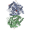
| |||||||||||||||||||||||||||||||||
|---|---|---|---|---|---|---|---|---|---|---|---|---|---|---|---|---|---|---|---|---|---|---|---|---|---|---|---|---|---|---|---|---|---|---|
| 1 | 
| |||||||||||||||||||||||||||||||||
| 2 | 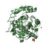
| |||||||||||||||||||||||||||||||||
| Unit cell |
| |||||||||||||||||||||||||||||||||
| Noncrystallographic symmetry (NCS) | NCS domain:
NCS domain segments: Ens-ID: ens_1 / End auth comp-ID: PRO / End label comp-ID: PRO
|
-Components
| #1: Protein | Mass: 36757.836 Da / Num. of mol.: 2 Source method: isolated from a genetically manipulated source Source: (gene. exp.) Verrucomicrobium spinosum (bacteria) / Production host: #2: Chemical | ChemComp-CU /   Mass: 63.546 Da / Num. of mol.: 11 / Source method: obtained synthetically / Formula: Cu / Feature type: SUBJECT OF INVESTIGATION Mass: 63.546 Da / Num. of mol.: 11 / Source method: obtained synthetically / Formula: Cu / Feature type: SUBJECT OF INVESTIGATION#3: Chemical |   Mass: 96.063 Da / Num. of mol.: 3 / Source method: obtained synthetically / Formula: SO4 Mass: 96.063 Da / Num. of mol.: 3 / Source method: obtained synthetically / Formula: SO4#4: Water | ChemComp-HOH / |  Mass: 18.015 Da / Num. of mol.: 688 / Source method: isolated from a natural source / Formula: H2O Mass: 18.015 Da / Num. of mol.: 688 / Source method: isolated from a natural source / Formula: H2OHas ligand of interest | Y | |
|---|
-Experimental details
-Experiment
| Experiment | Method: X-RAY DIFFRACTION / Number of used crystals: 1 |
|---|
- Sample preparation
| Crystal | Density Matthews: 2.18 Å3/Da / Density % sol: 43.63 % |
|---|---|
| Crystal grow | Temperature: 293 K / Method: vapor diffusion Details: 0.1 M ammonium sulfate, 0.1 M MES pH 6.5, 30% w/v PEG5000MME |
-Data collection
| Diffraction | Mean temperature: 100 K / Serial crystal experiment: N |
|---|---|
| Diffraction source | Source: SYNCHROTRON / Site: Diamond  / Beamline: I03 / Wavelength: 0.97626 Å / Beamline: I03 / Wavelength: 0.97626 Å |
| Detector | Type: DECTRIS EIGER2 X 16M / Detector: PIXEL / Date: Nov 26, 2021 |
| Radiation | Protocol: SINGLE WAVELENGTH / Monochromatic (M) / Laue (L): M / Scattering type: x-ray |
| Radiation wavelength | Wavelength: 0.97626 Å / Relative weight: 1 |
| Reflection | Resolution: 1.64→116.37 Å / Num. obs: 75699 / % possible obs: 98.2 % / Redundancy: 6.7 % / CC1/2: 0.998 / Net I/σ(I): 9.8 |
| Reflection shell | Resolution: 1.64→1.67 Å / Num. unique obs: 3073 / CC1/2: 0.362 |
- Processing
| Software |
| ||||||||||||||||||||||||||||||||||||||||||||||||||||||||||||
|---|---|---|---|---|---|---|---|---|---|---|---|---|---|---|---|---|---|---|---|---|---|---|---|---|---|---|---|---|---|---|---|---|---|---|---|---|---|---|---|---|---|---|---|---|---|---|---|---|---|---|---|---|---|---|---|---|---|---|---|---|---|
| Refinement | Method to determine structure: MOLECULAR REPLACEMENT Starting model: 4Z11, 4Z12, 5CE9 Resolution: 1.64→116.37 Å / Cor.coef. Fo:Fc: 0.967 / Cor.coef. Fo:Fc free: 0.955 / SU B: 2.723 / SU ML: 0.084 / Cross valid method: THROUGHOUT / σ(F): 0 / ESU R: 0.113 / ESU R Free: 0.109 / Stereochemistry target values: MAXIMUM LIKELIHOOD Details: HYDROGENS HAVE BEEN ADDED IN THE RIDING POSITIONS U VALUES : REFINED INDIVIDUALLY
| ||||||||||||||||||||||||||||||||||||||||||||||||||||||||||||
| Solvent computation | Ion probe radii: 0.8 Å / Shrinkage radii: 0.8 Å / VDW probe radii: 1.2 Å / Solvent model: MASK | ||||||||||||||||||||||||||||||||||||||||||||||||||||||||||||
| Displacement parameters | Biso max: 81.95 Å2 / Biso mean: 25.752 Å2 / Biso min: 10.7 Å2
| ||||||||||||||||||||||||||||||||||||||||||||||||||||||||||||
| Refinement step | Cycle: final / Resolution: 1.64→116.37 Å
| ||||||||||||||||||||||||||||||||||||||||||||||||||||||||||||
| Refine LS restraints |
| ||||||||||||||||||||||||||||||||||||||||||||||||||||||||||||
| Refine LS restraints NCS | Ens-ID: ens_1 / Number: 11706 / Refine-ID: X-RAY DIFFRACTION / Type: interatomic distance / Rms dev position: 0.05 Å / Weight position: 0.05
| ||||||||||||||||||||||||||||||||||||||||||||||||||||||||||||
| LS refinement shell | Resolution: 1.64→1.683 Å / Rfactor Rfree error: 0 / Total num. of bins used: 20
|
