 Movie
Movie Controller

[English] 日本語
 Yorodumi
- PDB-8bb8: Crystal structure of human aldehyde dehydrogenase ALDH3A1 in comp... -
+ Open data

- Basic information
| Entry | Database: PDB / ID: 8bb8 | ||||||
|---|---|---|---|---|---|---|---|
| Title | Crystal structure of human aldehyde dehydrogenase ALDH3A1 in complex with octanal | ||||||
 Components Components | Aldehyde dehydrogenase, dimeric NADP-preferring | ||||||
 Keywords Keywords | OXIDOREDUCTASE / aldehyde dehydrogenase / ALDH3A1 / odorant | ||||||
| Function / homology |  Function and homology information Function and homology informationaldehyde dehydrogenase [NAD(P)+] / 3-chloroallyl aldehyde dehydrogenase activity / benzaldehyde dehydrogenase (NAD+) activity / aldehyde dehydrogenase [NAD(P)+] activity / aldehyde metabolic process / alcohol dehydrogenase (NADP+) activity / aldehyde dehydrogenase (NAD+) activity / Phase I - Functionalization of compounds / xenobiotic metabolic process / lipid metabolic process ...aldehyde dehydrogenase [NAD(P)+] / 3-chloroallyl aldehyde dehydrogenase activity / benzaldehyde dehydrogenase (NAD+) activity / aldehyde dehydrogenase [NAD(P)+] activity / aldehyde metabolic process / alcohol dehydrogenase (NADP+) activity / aldehyde dehydrogenase (NAD+) activity / Phase I - Functionalization of compounds / xenobiotic metabolic process / lipid metabolic process / endoplasmic reticulum / : / plasma membrane / cytoplasm / cytosol Similarity search - Function | ||||||
| Biological species |  Homo sapiens (human) Homo sapiens (human) | ||||||
| Method |  X-RAY DIFFRACTION / SYNCHROTRON / MOLECULAR REPLACEMENT / Resolution: 1.8 Å X-RAY DIFFRACTION / SYNCHROTRON / MOLECULAR REPLACEMENT / Resolution: 1.8 Å | ||||||
 Authors Authors | Schwartz, M. / Neiers, F. | ||||||
| Funding support |  France, 1items France, 1items
| ||||||
 Citation Citation | Journal: Sci Rep / Year: 2023 Title: Characterization of human oxidoreductases involved in aldehyde odorant metabolism. Authors: Boichot, V. / Menetrier, F. / Saliou, J.M. / Lirussi, F. / Canon, F. / Folia, M. / Heydel, J.M. / Hummel, T. / Menzel, S. / Steinke, M. / Hackenberg, S. / Schwartz, M. / Neiers, F. | ||||||
| History |
|
- Structure visualization
| Structure viewer | Molecule: MolmilJmol/JSmol |
|---|
- Downloads & links
-Download
| PDBx/mmCIF format | 8bb8.cif.gz | 211.8 KB | Display | PDBx/mmCIF format |
|---|---|---|---|---|
| PDB format | pdb8bb8.ent.gz | 162.5 KB | Display | PDB format |
| PDBx/mmJSON format | 8bb8.json.gz | Tree view | PDBx/mmJSON format | |
| Others |  Other downloads Other downloads |
-Validation report
| Arichive directory | https://data.pdbj.org/pub/pdb/validation_reports/bb/8bb8ftp://data.pdbj.org/pub/pdb/validation_reports/bb/8bb8 | HTTPS FTP |
|---|
-Related structure data
| Related structure data | 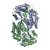 3szaS S: Starting model for refinement |
|---|---|
| Similar structure data |
-Links
 PDBj
PDBj

- Assembly
| Deposited unit | 
| ||||||||||||
|---|---|---|---|---|---|---|---|---|---|---|---|---|---|
| 1 |
| ||||||||||||
| Unit cell |
|
-Components
-Protein , 1 types, 2 molecules AB
| #1: Protein | Mass: 52089.445 Da / Num. of mol.: 2 Source method: isolated from a genetically manipulated source Source: (gene. exp.) Homo sapiens (human) / Gene: ALDH3A1, ALDH3 / Production host:  References: UniProt: P30838, aldehyde dehydrogenase [NAD(P)+] |
|---|
-Non-polymers , 5 types, 867 molecules 


| #2: Chemical | ChemComp-K /  Mass: 39.098 Da / Num. of mol.: 4 / Source method: obtained synthetically / Formula: K Mass: 39.098 Da / Num. of mol.: 4 / Source method: obtained synthetically / Formula: K#3: Chemical |  Mass: 59.044 Da / Num. of mol.: 2 / Source method: obtained synthetically / Formula: C2H3O2 Mass: 59.044 Da / Num. of mol.: 2 / Source method: obtained synthetically / Formula: C2H3O2#4: Chemical |  Mass: 128.212 Da / Num. of mol.: 2 / Source method: obtained synthetically / Formula: C8H16O Mass: 128.212 Da / Num. of mol.: 2 / Source method: obtained synthetically / Formula: C8H16O#5: Chemical | ChemComp-GOL / |  Mass: 92.094 Da / Num. of mol.: 1 / Source method: obtained synthetically / Formula: C3H8O3 Mass: 92.094 Da / Num. of mol.: 1 / Source method: obtained synthetically / Formula: C3H8O3#6: Water | ChemComp-HOH / | Mass: 18.015 Da / Num. of mol.: 858 / Source method: isolated from a natural source / Formula: H2O |
|---|
-Details
| Has ligand of interest | Y |
|---|
-Experimental details
-Experiment
| Experiment | Method: X-RAY DIFFRACTION / Number of used crystals: 1 |
|---|
- Sample preparation
| Crystal | Density Matthews: 2.14 Å3/Da / Density % sol: 42.46 % |
|---|---|
| Crystal grow | Temperature: 293 K / Method: vapor diffusion, sitting drop Details: 18 % PEG 3350, in 0.1 M potassium acetate pH 7.5 buffer |
-Data collection
| Diffraction | Mean temperature: 100 K / Serial crystal experiment: N |
|---|---|
| Diffraction source | Source: SYNCHROTRON / Site: SOLEIL / Beamline: PROXIMA 1 / Wavelength: 0.978565 Å |
| Detector | Type: DECTRIS EIGER X 16M / Detector: PIXEL / Date: Jun 3, 2021 |
| Radiation | Protocol: SINGLE WAVELENGTH / Monochromatic (M) / Laue (L): M / Scattering type: x-ray |
| Radiation wavelength | Wavelength: 0.978565 Å / Relative weight: 1 |
| Reflection | Resolution: 1.8→49.66 Å / Num. obs: 82079 / % possible obs: 97.6 % / Redundancy: 12.6 % / Biso Wilson estimate: 22.85 Å2 / CC1/2: 0.99 / Rmerge(I) obs: 0.318 / Rpim(I) all: 0.091 / Rrim(I) all: 0.331 / Net I/σ(I): 8.8 |
| Reflection shell | Resolution: 1.8→1.83 Å / Redundancy: 7.5 % / Mean I/σ(I) obs: 1.1 / Num. unique obs: 3334 / CC1/2: 0.541 / Rpim(I) all: 0.632 / % possible all: 74.4 |
- Processing
| Software |
| |||||||||||||||||||||||||||||||||||||||||||||||||||||||||||||||||||||||||||||||||||||||||||||||||||||||||||||||||||||||||||||||||||||||||||||||||||||||||||||||||||||||||||||||||||||||||||||||||||||||||||||||||||||||||
|---|---|---|---|---|---|---|---|---|---|---|---|---|---|---|---|---|---|---|---|---|---|---|---|---|---|---|---|---|---|---|---|---|---|---|---|---|---|---|---|---|---|---|---|---|---|---|---|---|---|---|---|---|---|---|---|---|---|---|---|---|---|---|---|---|---|---|---|---|---|---|---|---|---|---|---|---|---|---|---|---|---|---|---|---|---|---|---|---|---|---|---|---|---|---|---|---|---|---|---|---|---|---|---|---|---|---|---|---|---|---|---|---|---|---|---|---|---|---|---|---|---|---|---|---|---|---|---|---|---|---|---|---|---|---|---|---|---|---|---|---|---|---|---|---|---|---|---|---|---|---|---|---|---|---|---|---|---|---|---|---|---|---|---|---|---|---|---|---|---|---|---|---|---|---|---|---|---|---|---|---|---|---|---|---|---|---|---|---|---|---|---|---|---|---|---|---|---|---|---|---|---|---|---|---|---|---|---|---|---|---|---|---|---|---|---|---|---|---|
| Refinement | Method to determine structure: MOLECULAR REPLACEMENT Starting model: 3SZA Resolution: 1.8→49.66 Å / SU ML: 0.2112 / Cross valid method: FREE R-VALUE / σ(F): 1.34 / Phase error: 23.1454 Stereochemistry target values: GeoStd + Monomer Library + CDL v1.2
| |||||||||||||||||||||||||||||||||||||||||||||||||||||||||||||||||||||||||||||||||||||||||||||||||||||||||||||||||||||||||||||||||||||||||||||||||||||||||||||||||||||||||||||||||||||||||||||||||||||||||||||||||||||||||
| Solvent computation | Shrinkage radii: 0.9 Å / VDW probe radii: 1.11 Å / Solvent model: FLAT BULK SOLVENT MODEL | |||||||||||||||||||||||||||||||||||||||||||||||||||||||||||||||||||||||||||||||||||||||||||||||||||||||||||||||||||||||||||||||||||||||||||||||||||||||||||||||||||||||||||||||||||||||||||||||||||||||||||||||||||||||||
| Displacement parameters | Biso mean: 26.32 Å2 | |||||||||||||||||||||||||||||||||||||||||||||||||||||||||||||||||||||||||||||||||||||||||||||||||||||||||||||||||||||||||||||||||||||||||||||||||||||||||||||||||||||||||||||||||||||||||||||||||||||||||||||||||||||||||
| Refinement step | Cycle: LAST / Resolution: 1.8→49.66 Å
| |||||||||||||||||||||||||||||||||||||||||||||||||||||||||||||||||||||||||||||||||||||||||||||||||||||||||||||||||||||||||||||||||||||||||||||||||||||||||||||||||||||||||||||||||||||||||||||||||||||||||||||||||||||||||
| Refine LS restraints |
| |||||||||||||||||||||||||||||||||||||||||||||||||||||||||||||||||||||||||||||||||||||||||||||||||||||||||||||||||||||||||||||||||||||||||||||||||||||||||||||||||||||||||||||||||||||||||||||||||||||||||||||||||||||||||
| LS refinement shell |
|
